WB
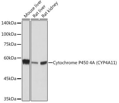
Western blot analysis of various lysates using Cytochrome P450 4A (CYP4A11) Rabbit mAb at 1:1000 dilution. Secondary antibody: HRP-conjugated Goat anti-Rabbit IgG (H+L)at 1:10000 dilution. Lysates/proteins: 25μg per lane. Blocking buffer: 3% nonfat dry milk in TBST. Detection: ECL Basic Kit. Exposure time: 10s.IHC
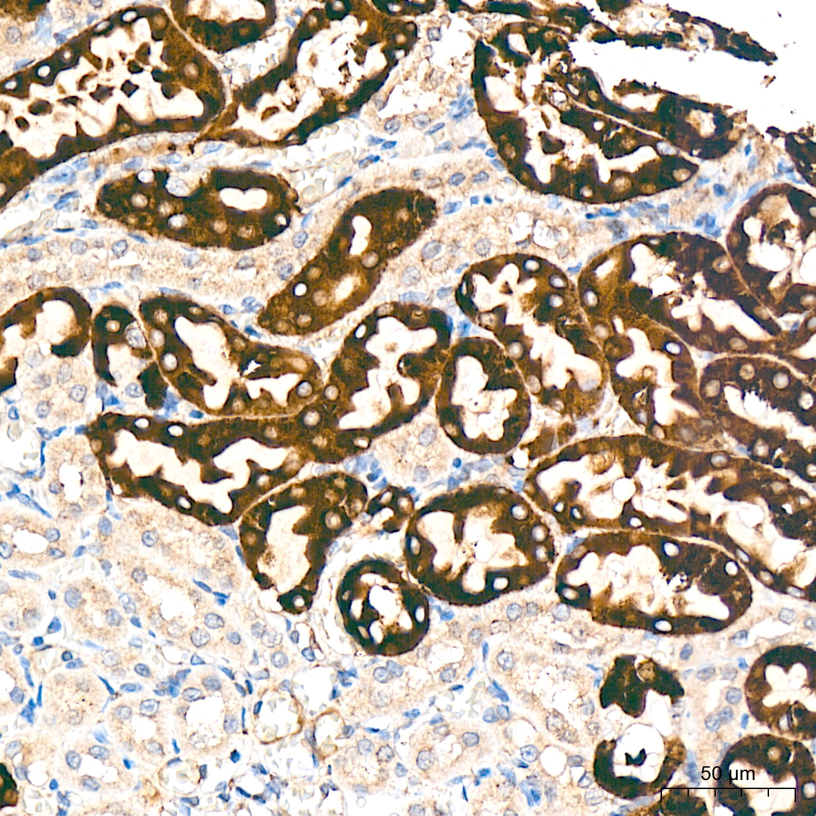
Immunohistochemistry analysis of paraffin embedded Rat kidney tissue using Cytochrome P450 4A (CYP4A11) Rabbit mAb at a dilution of 1:200 (40x lens). High pressure antigen retrieval was performed with 0.01 M citrate buffer (pH 6.0) prior to IHC staining.IHC
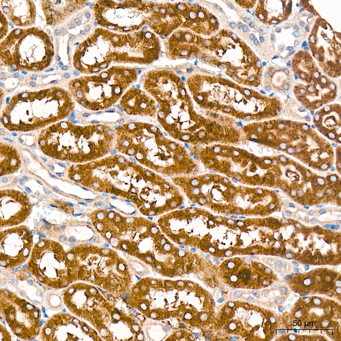
Immunohistochemistry analysis of paraffin embedded Mouse kidney tissue using Cytochrome P450 4A (CYP4A11) Rabbit mAb at a dilution of 1:200 (40x lens). High pressure antigen retrieval was performed with 0.01 M citrate buffer (pH 6.0) prior to IHC staining.IHC
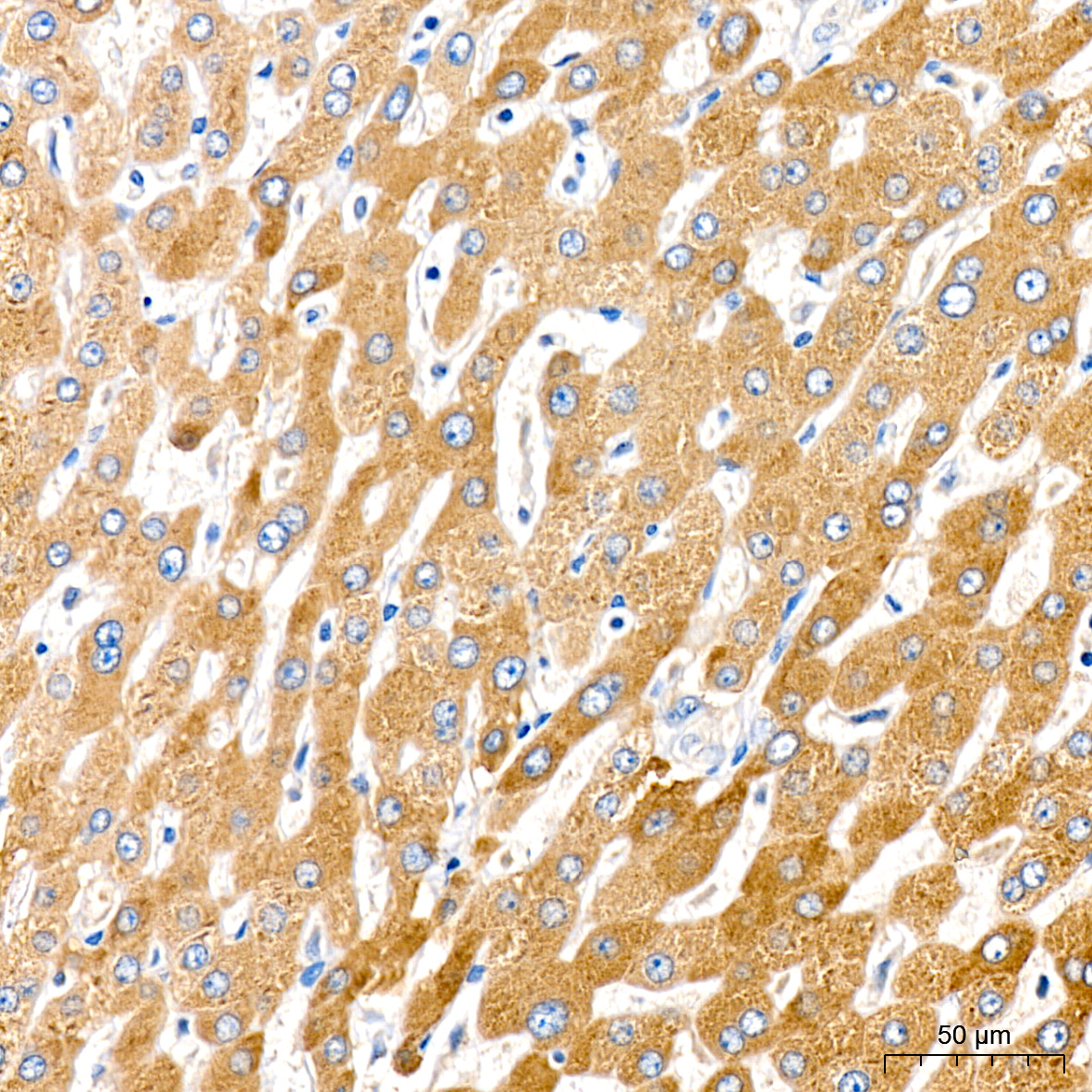
Immunohistochemistry analysis of paraffin embedded Human liver tissue using Cytochrome P450 4A (CYP4A11) Rabbit mAb at a dilution of 1:200 (40x lens). High pressure antigen retrieval was performed with 0.01 M citrate buffer (pH 6.0) prior to IHC staining.| Product Name | Cytochrome P450 4A (CYP4A11) Rabbit mAb |
|---|---|
| Antibody Type | Primary Antibodies |
| Immunogen | Synthetic peptide. |
| Clonality | Monoclonal |
|---|---|
| Isotype | IgG |
| Host Species | Rabbit |
| Tested Applications | IHCWB |
| WB:1:500-1:2000 IHC:1:50-1:200 |
|
| Species Reactivity | HumanMouseRat |
| Concentration | 1mg/ml |
| Purification | Affinity purified |
| Gene Symbol | CYP4A11 |
|---|---|
| Gene Synonyms | CP4Y CYP4A2 CYP4AII CYPIVA11 |
| Gene Full Name | cytochrome P450 family 4 subfamily A member 11 |
| Gene Summary | This gene encodes a member of the cytochrome P450 superfamily of enzymes. The cytochrome P450 proteins are monooxygenases which catalyze many reactions involved in drug metabolism and synthesis of cholesterol, steroids and other lipids. This protein localizes to the endoplasmic reticulum and hydroxylates medium-chain fatty acids such as laurate and myristate. Multiple transcript variants have been found for this gene. [provided by RefSeq, Jan 2016] |
| Alternative Names | CP4Y CYP4A2 CYP4AII CYPIVA11 Cytochrome P450 4A (CYP4A11) |
| Molecular Weight(MW) | 59kDa |
| Cellular Localization | Apical plasma membrane, Cytoplasm. |
WB
Western blot analysis of various lysates using Cytochrome P450 4A (CYP4A11) Rabbit mAb at 1:1000 dilution. Secondary antibody: HRP-conjugated Goat anti-Rabbit IgG (H+L)at 1:10000 dilution. Lysates/proteins: 25μg per lane. Blocking buffer: 3% nonfat dry milk in TBST. Detection: ECL Basic Kit. Exposure time: 10s.
IHC
Immunohistochemistry analysis of paraffin embedded Rat kidney tissue using Cytochrome P450 4A (CYP4A11) Rabbit mAb at a dilution of 1:200 (40x lens). High pressure antigen retrieval was performed with 0.01 M citrate buffer (pH 6.0) prior to IHC staining.
IHC
Immunohistochemistry analysis of paraffin embedded Mouse kidney tissue using Cytochrome P450 4A (CYP4A11) Rabbit mAb at a dilution of 1:200 (40x lens). High pressure antigen retrieval was performed with 0.01 M citrate buffer (pH 6.0) prior to IHC staining.
IHC
Immunohistochemistry analysis of paraffin embedded Human liver tissue using Cytochrome P450 4A (CYP4A11) Rabbit mAb at a dilution of 1:200 (40x lens). High pressure antigen retrieval was performed with 0.01 M citrate buffer (pH 6.0) prior to IHC staining.| Application Notes | WB:1:500-1:2000 IHC:1:50-1:200 |
|---|
| Form | Liquid |
|---|---|
| Storage Instructions | Store at -20℃. Avoid freeze / thaw cycles. |
| Storage Buffer | PBS with 0.09% Sodium azide, 0.05% BSA, 50% glycerol, pH7.3. |


