WB
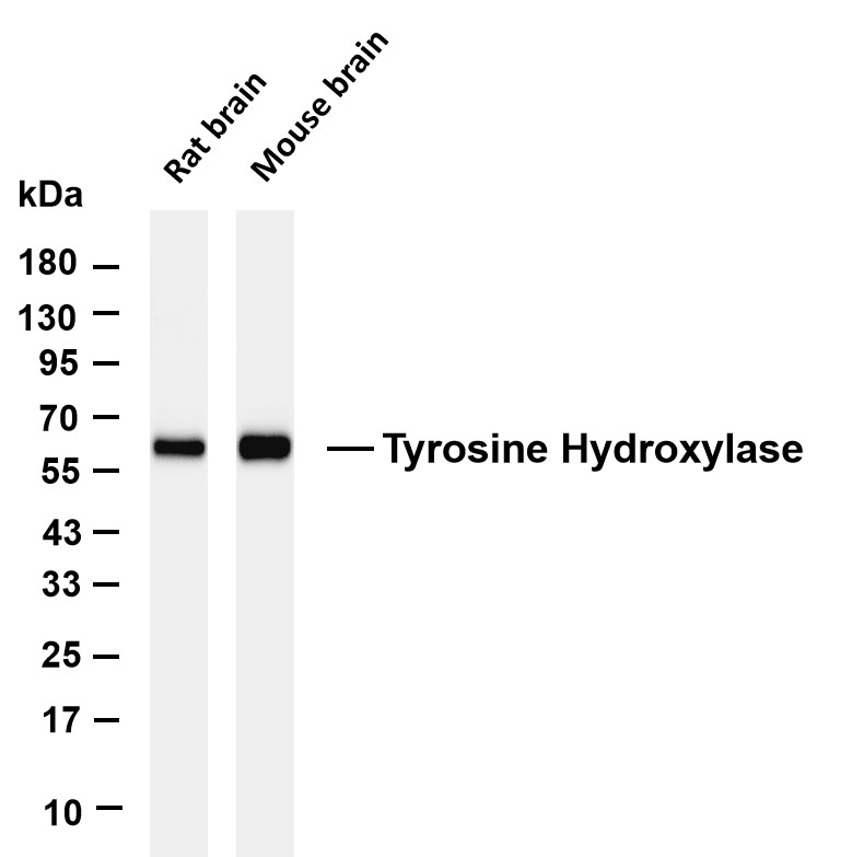
Various whole cell lysates were separated by 4-20% SDS-PAGE, and the membrane was blotted with anti-Tyrosine Hydroxylase antibody. The HRP-conjugated Goat anti-Rabbit IgG (H + L) antibody was used to detect the antibody. Lane 1: Rat brain Lane 2: Mouse brain Predicted band size: 59kDa Observed band size: 59kDaIHC
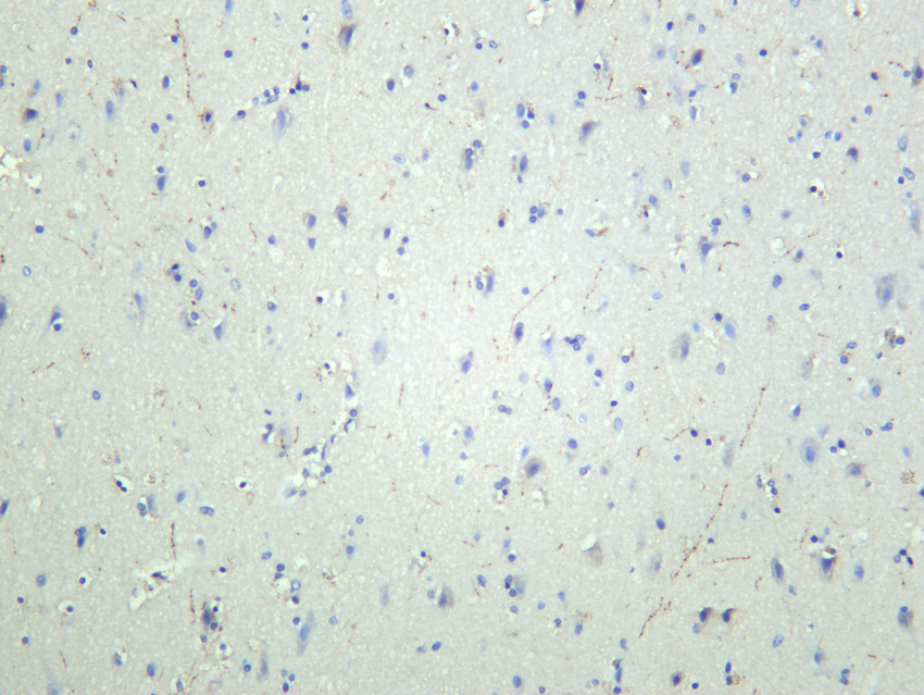
Human brain was stained with anti-Tyrosine Hydroxylase rabbit antibodyICC/IF
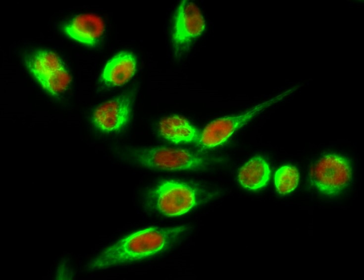
Immunofluorescence analysis of Hela cell. 1,TH Monoclonal Antibody(red) was diluted at 1:200(4° overnight). GAPDH Monoclonal Antibody(2B8)(green) was diluted at 1:200(4° overnight). 2, Goat Anti Rabbit Alexa Fluor 594 Catalog:RS3611 was diluted at 1:1000(room temperature, 50min). Goat Anti Mouse Alexa Fluor 488 was diluted at 1:1000(room temperature, 50min).| Product Name | Tyrosine Hydroxylase Rabbit mAb |
|---|---|
| Antibody Type | Primary Antibodies |
| Immunogen | Recombinant |
| Clonality | Monoclonal |
|---|---|
| Isotype | IgG |
| Host Species | Rabbit |
| Tested Applications | ELISAICC/IFIHCIPWB |
| WB:1:2000-1:10000 IHC:1:1000-1:4000 ICC:1:200-1:1000 IP:1:50-1:200 ICC/IF:1:50-1:200 ELISA:1:1000-1:10000 |
|
| Species Reactivity | HumanMouseRat |
| Concentration | 1mg/ml |
| Purification | Protein A |
| Gene Symbol | TH |
|---|---|
| Gene Synonyms | TYH DYT14 DYT5b |
| Gene Full Name | tyrosine hydroxylase |
| Gene Summary | The protein encoded by this gene is involved in the conversion of tyrosine to dopamine. It is the rate-limiting enzyme in the synthesis of catecholamines, hence plays a key role in the physiology of adrenergic neurons. Mutations in this gene have been associated with autosomal recessive Segawa syndrome. Alternatively spliced transcript variants encoding different isoforms have been noted for this gene. [provided by RefSeq, Jul 2008] |
| Alternative Names | Tyrosine 3-monooxygenase Tyrosine 3-hydroxylase TH |
| Molecular Weight(MW) | 59kD |
| Cellular Localization | #Cell projection #Cytoplasm #Cytoplasmic vesicle #Nucleus #Synapse |
WB
Various whole cell lysates were separated by 4-20% SDS-PAGE, and the membrane was blotted with anti-Tyrosine Hydroxylase antibody. The HRP-conjugated Goat anti-Rabbit IgG (H + L) antibody was used to detect the antibody. Lane 1: Rat brain Lane 2: Mouse brain Predicted band size: 59kDa Observed band size: 59kDa
IHC
Human brain was stained with anti-Tyrosine Hydroxylase rabbit antibody
ICC/IF
Immunofluorescence analysis of Hela cell. 1,TH Monoclonal Antibody(red) was diluted at 1:200(4° overnight). GAPDH Monoclonal Antibody(2B8)(green) was diluted at 1:200(4° overnight). 2, Goat Anti Rabbit Alexa Fluor 594 Catalog:RS3611 was diluted at 1:1000(room temperature, 50min). Goat Anti Mouse Alexa Fluor 488 was diluted at 1:1000(room temperature, 50min).| Application Notes | WB:1:2000-1:10000 IHC:1:1000-1:4000 ICC:1:200-1:1000 IP:1:50-1:200 ICC/IF:1:50-1:200 ELISA:1:1000-1:10000 |
|---|
| Form | Liquid |
|---|---|
| Storage Instructions | -15°C to -25°C/1 year(Do not lower than -25°C. |
| Storage Buffer | PBS, 50% glycerol, 0.05% Proclin 300, 0.05%BSA. |


