WB
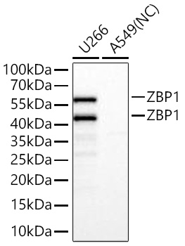
Western blot analysis of various lysates using ZBP1 Rabbit mAb at 1:11000 dilution incubated overnight at 4℃. Secondary antibody: HRP-conjugated Goat anti-Rabbit IgG (H+L) at 1:10000 dilution. Lysates/proteins: 25 μg per lane. Blocking buffer: 3% nonfat dry milk in TBST. Detection: ECL Basic Kit. Negative control (NC): A549 Exposure time: 1s.IHC
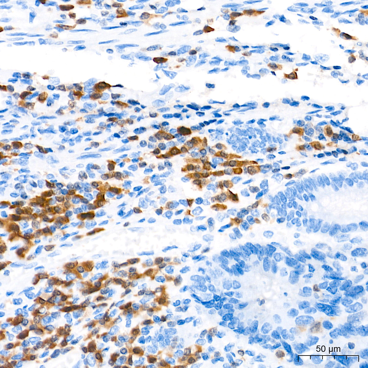
Immunohistochemistry analysis of paraffin embedded Human colon carcinoma tissue using ZBP1 Rabbit mAb at a dilution of 1:500 (40x lens). High pressure antigen retrieval performed with 0.01M Tris EDTA Buffer (pH 9.0) prior to IHC staining.IHC
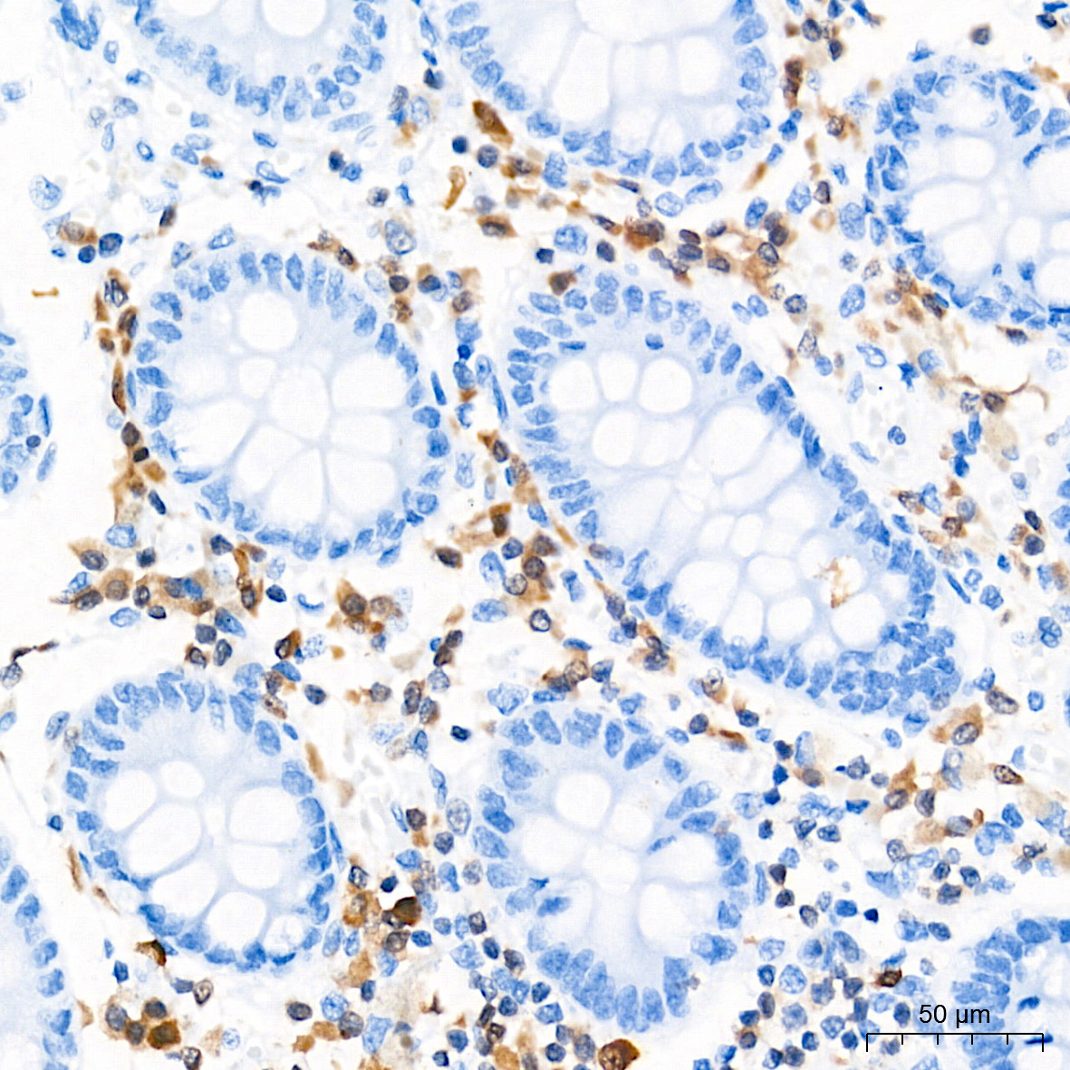
Immunohistochemistry analysis of paraffin embedded Human colon tissue using ZBP1 Rabbit mAb at a dilution of 1:500 (40x lens). High pressure antigen retrieval performed with 0.01M Tris-EDTA Buffer (pH 9.0) prior to IHC staining.ICC/IF
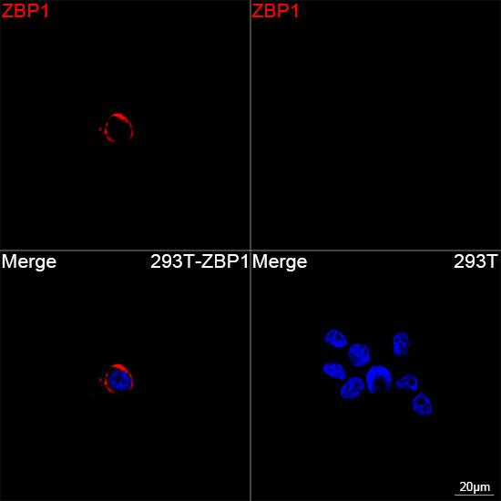
Confocal imaging of 293T cells transfected with ZBP1 using ZBP1 Rabbit mAb (dilution 1:200) followed by a further incubation with Cy3 Goat Anti-Rabbit IgG (H+L) (dilution 1:500) (Red). DAPI was used for nuclear staining (Blue). Objective: 100x.IP
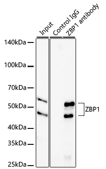
Immunoprecipitation of ZBP1 from 600 µg extracts of U266 cells was performed using 1 µg of ZBP1 Rabbit mAb. Rabbit IgG isotype control was used to precipitate the Control IgG sample. IP samples were eluted with 1X non-reducing Laemmli Buffer. The Input lane represents 10% of the total input. Western blot analysis of immunoprecipitates was conducted using ZBP1 Rabbit mAb at a dilution of 1:3000.| Product Name | ZBP1 Rabbit mAb |
|---|---|
| Antibody Type | Primary Antibodies |
| Immunogen | Recombinant protein. |
| Clonality | Monoclonal |
|---|---|
| Isotype | IgG |
| Host Species | Rabbit |
| Tested Applications | ICC/IFIHCIPWB |
| WB:1:5000-1:20000 IHC:1:200-1:800 ICC/IF:1:100-1:400 IP:0.5μg-4μg antibody for 500μg-700μg extracts of whole cells |
|
| Species Reactivity | Human |
| Concentration | 1mg/ml |
| Purification | Affinity purified |
| Gene Symbol | ZBP1 |
|---|---|
| Gene Synonyms | DAI DLM1 DLM-1 C20orf183 |
| Gene Full Name | Z-DNA binding protein 1 |
| Gene Summary | This gene encodes a Z-DNA binding protein. The encoded protein plays a role in the innate immune response by binding to foreign DNA and inducing type-I interferon production. Alternatively spliced transcript variants encoding multiple isoforms have been observed for this gene. [provided by RefSeq, Dec 2011] |
| Alternative Names | DAI DLM1 DLM-1 C20orf183 |
| Molecular Weight(MW) | 44kDa/58kDa |
| Cellular Localization | cytoplasm, cytosol, nucleus. |
WB
Western blot analysis of various lysates using ZBP1 Rabbit mAb at 1:11000 dilution incubated overnight at 4℃. Secondary antibody: HRP-conjugated Goat anti-Rabbit IgG (H+L) at 1:10000 dilution. Lysates/proteins: 25 μg per lane. Blocking buffer: 3% nonfat dry milk in TBST. Detection: ECL Basic Kit. Negative control (NC): A549 Exposure time: 1s.
IHC
Immunohistochemistry analysis of paraffin embedded Human colon carcinoma tissue using ZBP1 Rabbit mAb at a dilution of 1:500 (40x lens). High pressure antigen retrieval performed with 0.01M Tris EDTA Buffer (pH 9.0) prior to IHC staining.
IHC
Immunohistochemistry analysis of paraffin embedded Human colon tissue using ZBP1 Rabbit mAb at a dilution of 1:500 (40x lens). High pressure antigen retrieval performed with 0.01M Tris-EDTA Buffer (pH 9.0) prior to IHC staining.
ICC/IF
Confocal imaging of 293T cells transfected with ZBP1 using ZBP1 Rabbit mAb (dilution 1:200) followed by a further incubation with Cy3 Goat Anti-Rabbit IgG (H+L) (dilution 1:500) (Red). DAPI was used for nuclear staining (Blue). Objective: 100x.
IP
Immunoprecipitation of ZBP1 from 600 µg extracts of U266 cells was performed using 1 µg of ZBP1 Rabbit mAb. Rabbit IgG isotype control was used to precipitate the Control IgG sample. IP samples were eluted with 1X non-reducing Laemmli Buffer. The Input lane represents 10% of the total input. Western blot analysis of immunoprecipitates was conducted using ZBP1 Rabbit mAb at a dilution of 1:3000.| Application Notes | WB:1:5000-1:20000 IHC:1:200-1:800 ICC/IF:1:100-1:400 IP:0.5μg-4μg antibody for 500μg-700μg extracts of whole cells |
|---|
| Form | Liquid |
|---|---|
| Storage Instructions | Store at -20℃. Avoid freeze / thaw cycles. |
| Storage Buffer | PBS with 0.09% Sodium azide, 0.05% BSA, 50% glycerol, pH7.3. |


