WB
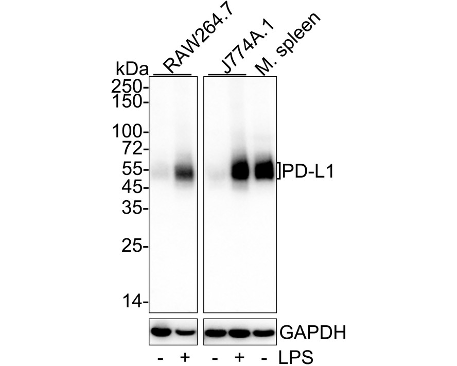
Western blot analysis of PD-L1 on different lysates with Rabbit anti-PD-L1 antibody at 1/2,000 dilution. Lane 1: RAW264.7 cell lysate, Lane 2: RAW264.7 treated with 10μg/mL LPS for 8 hours cell lysate, Lane 3: J774A.1 cell lysate, Lane 4: J774A.1 treated with 1μg/mL LPS for 24 hours cell lysate, Lane 5: Mouse spleen tissue lysate, Lysates/proteins at 30 µg/Lane. Exposure time: 46 seconds; 4-20% SDS-PAGE gel. Proteins were transferred to a PVDF membrane and blocked with 5% NFDM/TBST for 1 hour at room temperature. The primary antibody at 1/2,000 dilution was used in 5% NFDM/TBST at 4℃ overnight. Goat Anti-Rabbit IgG - HRP Secondary Antibody at 1/50,000 dilution was used for 1 hour at room temperature.IHC
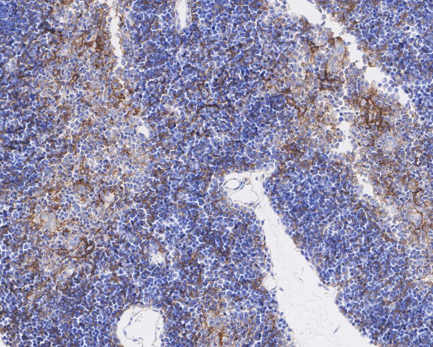
Immunohistochemical analysis of paraffin-embedded mouse thymus tissue with Rabbit anti-PD-L1 antibody at 1/200 dilution. The section was pre-treated using heat mediated antigen retrieval with Tris-EDTA buffer (pH 9.0) for 20 minutes. The tissues were blocked in 1% BSA for 20 minutes at room temperature, washed with ddH2O and PBS, and then probed with the primary antibody at 1/200 dilution for 1 hour at room temperature. The detection was performed using an HRP conjugated compact polymer system. DAB was used as the chromogen. Tissues were counterstained with hematoxylin and mounted with DPX.IHC
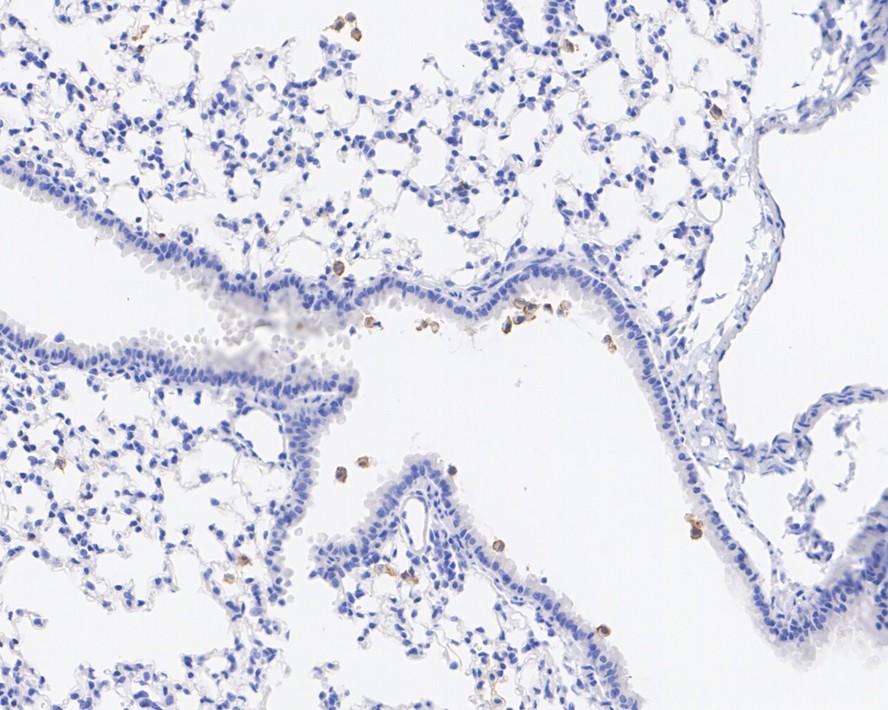
Immunohistochemical analysis of paraffin-embedded mouse lung tissue with Rabbit anti-PD-L1 antibody at 1/200 dilution. The section was pre-treated using heat mediated antigen retrieval with Tris-EDTA buffer (pH 9.0) for 20 minutes. The tissues were blocked in 1% BSA for 20 minutes at room temperature, washed with ddH2O and PBS, and then probed with the primary antibody at 1/200 dilution for 1 hour at room temperature. The detection was performed using an HRP conjugated compact polymer system. DAB was used as the chromogen. Tissues were counterstained with hematoxylin and mounted with DPX.mIHC
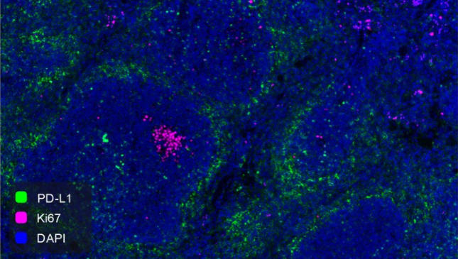
Fluorescence multiplex immunohistochemical analysis of mouse spleen (Formalin/PFA-fixed paraffin-embedded sections). Panel A: the merged image of anti-PD-L1 (Green) and anti-Ki67 (Violet) on spleen. HRP Conjugated UltraPolymer Goat Polyclonal Antibody Mouse/Rabbit was used as a secondary antibody. The immunostaining was performed with Three-Fluorescence immunohistochemical mouse/rabbit kit. The section was incubated in two rounds of staining: in the order of anti-PD-L1 (1/500 dilution) and anti-Ki67 (1/2,000 dilution) for 20 mins at room temperature. Each round was followed by a separate fluorescent tyramide signal amplification system. Heat mediated antigen retrieval with Tris-EDTA buffer (pH 9.0) for 30 mins at 95℃. DAPI (blue) was used as a nuclear counter stain. Image acquisition was performed with Olympus VS200 Slide Scanner.| Product Name | PD-L1 Recombinant Rabbit Monoclonal Antibody |
|---|---|
| Antibody Type | Primary Antibodies |
| Immunogen | Recombinant protein within mouse PD-L1 aa 1-250 / 290. |
| Clonality | monoclonal |
|---|---|
| Isotype | IgG |
| Host Species | Rabbit |
| Tested Applications | IHCWBmIHC |
| WB:1:2000 IHC:1:200 mIHC:1:500 |
|
| Species Reactivity | Mouse |
| Concentration | 1mg/ml |
| Purification | Protein A |
| Gene Symbol | Cd274 |
|---|---|
| Gene Synonyms | B7h1 Pdl1 Pdcd1l1 Pdcd1lg1 A530045L16Rik |
| Gene Full Name | CD274 antigen |
| Gene Summary | The protein encoded by this gene is an immune inhibitory receptor ligand that is expressed by hematopoietic and non-hematopoietic cells, such as T cells and B cells and various types of tumor cells. The encoded protein is a type I transmembrane protein that has immunoglobulin V-like and C-like domains. Interaction of this ligand with its receptor inhibits T-cell activation and cytokine production. During infection or inflammation of normal tissue, this interaction is important for preventing autoimmunity by maintaining homeostasis of the immune response. In tumor microenvironments, this interaction provides an immune escape for tumor cells through cytotoxic T-cell inactivation. Mice deficient for this gene display a variety of phenotypes including decreased allogeneic fetal survival rates and severe experimental autoimmune encephalomyelitis. [provided by RefSeq, Sep 2015] |
| Alternative Names | B7 H antibody B7 H1 antibody B7 homolog 1 antibody B7-H1 antibody B7H antibody B7H1 antibody CD 274 antibody CD-274 antibody CD274 antibody CD274 antigen antibody B7 H antibody B7 H1 antibody B7 homolog 1 antibody B7-H1 antibody B7H antibody B7H1 antibody CD 274 antibody CD-274 antibody CD274 antibody CD274 antigen antibody CD274 molecule antibody MGC142294 antibody MGC142296 antibody OTTHUMP00000021029 antibody PD L1 antibody PD-L1 antibody PD1L1_HUMAN antibody PDCD1 ligand 1 antibody PDCD1L1 antibody PDCD1LG1 antibody PDL 1 antibody PDL1 antibody Programmed cell death 1 ligand 1 antibody Programmed death ligand 1 antibody RGD1566211 antibody |
| Molecular Weight(MW) | 33kDa(Observed band size: 45-60kDa) |
| Cellular Localization | Cell membrane, Early endosome membrane, Recycling endosome membrane. |
WB
Western blot analysis of PD-L1 on different lysates with Rabbit anti-PD-L1 antibody at 1/2,000 dilution. Lane 1: RAW264.7 cell lysate, Lane 2: RAW264.7 treated with 10μg/mL LPS for 8 hours cell lysate, Lane 3: J774A.1 cell lysate, Lane 4: J774A.1 treated with 1μg/mL LPS for 24 hours cell lysate, Lane 5: Mouse spleen tissue lysate, Lysates/proteins at 30 µg/Lane. Exposure time: 46 seconds; 4-20% SDS-PAGE gel. Proteins were transferred to a PVDF membrane and blocked with 5% NFDM/TBST for 1 hour at room temperature. The primary antibody at 1/2,000 dilution was used in 5% NFDM/TBST at 4℃ overnight. Goat Anti-Rabbit IgG - HRP Secondary Antibody at 1/50,000 dilution was used for 1 hour at room temperature.
IHC
Immunohistochemical analysis of paraffin-embedded mouse thymus tissue with Rabbit anti-PD-L1 antibody at 1/200 dilution. The section was pre-treated using heat mediated antigen retrieval with Tris-EDTA buffer (pH 9.0) for 20 minutes. The tissues were blocked in 1% BSA for 20 minutes at room temperature, washed with ddH2O and PBS, and then probed with the primary antibody at 1/200 dilution for 1 hour at room temperature. The detection was performed using an HRP conjugated compact polymer system. DAB was used as the chromogen. Tissues were counterstained with hematoxylin and mounted with DPX.
IHC
Immunohistochemical analysis of paraffin-embedded mouse lung tissue with Rabbit anti-PD-L1 antibody at 1/200 dilution. The section was pre-treated using heat mediated antigen retrieval with Tris-EDTA buffer (pH 9.0) for 20 minutes. The tissues were blocked in 1% BSA for 20 minutes at room temperature, washed with ddH2O and PBS, and then probed with the primary antibody at 1/200 dilution for 1 hour at room temperature. The detection was performed using an HRP conjugated compact polymer system. DAB was used as the chromogen. Tissues were counterstained with hematoxylin and mounted with DPX.
mIHC
Fluorescence multiplex immunohistochemical analysis of mouse spleen (Formalin/PFA-fixed paraffin-embedded sections). Panel A: the merged image of anti-PD-L1 (Green) and anti-Ki67 (Violet) on spleen. HRP Conjugated UltraPolymer Goat Polyclonal Antibody Mouse/Rabbit was used as a secondary antibody. The immunostaining was performed with Three-Fluorescence immunohistochemical mouse/rabbit kit. The section was incubated in two rounds of staining: in the order of anti-PD-L1 (1/500 dilution) and anti-Ki67 (1/2,000 dilution) for 20 mins at room temperature. Each round was followed by a separate fluorescent tyramide signal amplification system. Heat mediated antigen retrieval with Tris-EDTA buffer (pH 9.0) for 30 mins at 95℃. DAPI (blue) was used as a nuclear counter stain. Image acquisition was performed with Olympus VS200 Slide Scanner.| Application Notes | WB:1:2000 IHC:1:200 mIHC:1:500 |
|---|
| Form | Liquid |
|---|---|
| Storage Instructions | Store at +4℃ after thawing. Aliquot store at -20℃. Avoid repeated freeze / thaw cycles. |
| Storage Buffer | PBS (pH7.4), 0.1% BSA, 40% Glycerol. Preservative: 0.05% Sodium Azide. |


