WB
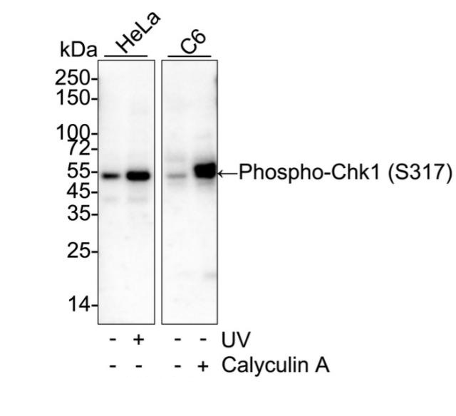
Western blot analysis of Phospho-Chk1 (S317) on different lysates with Rabbit anti-Phospho-Chk1 (S317) antibody at 1/1,000 dilution. Lane 1: HeLa cell lysate, Lane 2: HeLa treated with UV for 1 hour cell lysate, Lane 3: C6 cell lysate, Lane 4: C6 treated with 100nM Calyculin A for 30 minutes cell lysate, Lysates/proteins at 20 µg/Lane. Exposure time: 43 seconds; 4-20% SDS-PAGE gel. Proteins were transferred to a PVDF membrane and blocked with 5% NFDM/TBST for 1 hour at room temperature. The primary antibody at 1/1,000 dilution was used in 5% NFDM/TBST at 4℃ overnight. Goat Anti-Rabbit IgG - HRP Secondary Antibody at 1/50,000 dilution was used for 1 hour at room temperature.WB
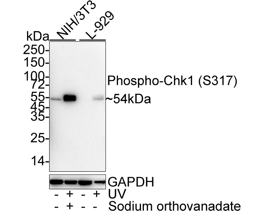
Western blot analysis of Phospho-Chk1 (S317) on different lysates with Rabbit anti-Phospho-Chk1 (S317) antibody at 1/1,000 dilution. Lane 1: NIH/3T3 cell lysate, Lane 2: NIH/3T3 treated with UV for 40 minutes add 1mM Sodium orthovanadate and recovery for 30 minutes cell lysate, Lane 3: L-929 cell lysate, Lane 4: L-929 treated with UV for 3 hours cell lysate, Lysates/proteins at 20 µg/Lane. Exposure time: 1 minute; 4-20% SDS-PAGE gel. Proteins were transferred to a PVDF membrane and blocked with 5% NFDM/TBST for 1 hour at room temperature. The primary antibody at 1/1,000 dilution was used in 5% NFDM/TBST at 4℃ overnight. Goat Anti-Rabbit IgG - HRP Secondary Antibody at 1/50,000 dilution was used for 1 hour at room temperature.IHC
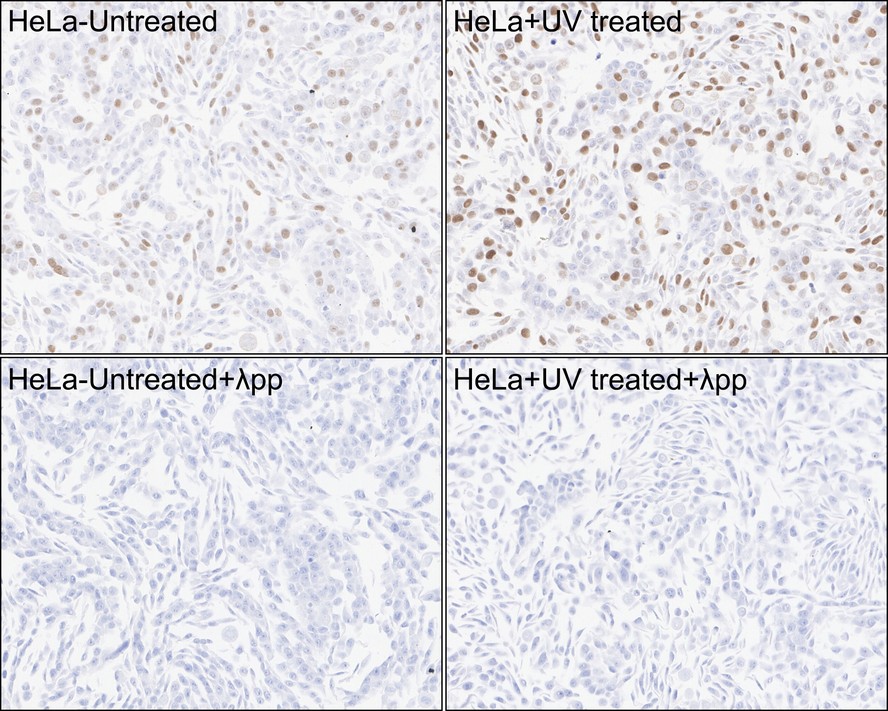
Immunohistochemical analysis of paraffin-embedded HeLa cells treated with or without UV for 40 minutes with Rabbit anti-Phospho-Chk1 (S317) antibody at 1/1,000 dilution. The section was pre-treated using heat mediated antigen retrieval with sodium citrate buffer (pH 6.0) (high pressure) for 2 minutes. The tissues were blocked in 1% BSA for 20 minutes at room temperature, washed with ddH2O and PBS, and then probed with the primary antibody at 1/1,000 dilution for 1 hour at room temperature. The detection was performed using an HRP conjugated compact polymer system. DAB was used as the chromogen. Tissues were counterstained with hematoxylin and mounted with DPX.| Product Name | Phospho-Chk1 (S317) Recombinant Rabbit Monoclonal Antibody |
|---|---|
| Antibody Type | Primary Antibodies |
| Immunogen | Synthetic phospho-peptide corresponding to residues surrounding Ser317 of Human Chk1. |
| Modification | p-S317 |
| Clonality | monoclonal |
|---|---|
| Isotype | IgG |
| Host Species | Rabbit |
| Tested Applications | IHCWB |
| WB:1:1000 IHC:1:1000 |
|
| Species Reactivity | HumanMouseRat |
| Concentration | 1mg/ml |
| Purification | Protein A |
| Gene Symbol | CHEK1 |
|---|---|
| Gene Synonyms | CHK1 OZEMA21 |
| Gene Full Name | checkpoint kinase 1 |
| Gene Summary | The protein encoded by this gene belongs to the Ser/Thr protein kinase family. It is required for checkpoint mediated cell cycle arrest in response to DNA damage or the presence of unreplicated DNA. This protein acts to integrate signals from ATM and ATR, two cell cycle proteins involved in DNA damage responses, that also associate with chromatin in meiotic prophase I. Phosphorylation of CDC25A protein phosphatase by this protein is required for cells to delay cell cycle progression in response to double-strand DNA breaks. Several alternatively spliced transcript variants have been found for this gene. [provided by RefSeq, Oct 2011] |
| Molecular Weight(MW) | 54kDa |
| Function | Checkpoint kinases (Chks) are protein kinases that are involved in cell cycle control. Two checkpoint kinase subtypes have been identified, Chk1 and Chk2. Chk1 is a central component of genome surveillance pathways and is a key regulator of the cell cycle and cell survival. Chk1 is required for the initiation of DNA damage checkpoints and has recently been shown to play a role in the normal (unperturbed) cell cycle. Chk1 impacts various stages of the cell cycle including the S phase, G2/M transition and M phaase. In addition to mediating cell cycle checkpoints, Chk1 also contributes to DNA repair processes, gene transcription, egg production, embryo development, cellular responses to HIV infection and somatic cell viability. |
| Cellular Localization | Nucleus, Chromosome, Cytoplasm, cytoskeleton, microtubule organizing center, centrosome. |
WB
Western blot analysis of Phospho-Chk1 (S317) on different lysates with Rabbit anti-Phospho-Chk1 (S317) antibody at 1/1,000 dilution. Lane 1: HeLa cell lysate, Lane 2: HeLa treated with UV for 1 hour cell lysate, Lane 3: C6 cell lysate, Lane 4: C6 treated with 100nM Calyculin A for 30 minutes cell lysate, Lysates/proteins at 20 µg/Lane. Exposure time: 43 seconds; 4-20% SDS-PAGE gel. Proteins were transferred to a PVDF membrane and blocked with 5% NFDM/TBST for 1 hour at room temperature. The primary antibody at 1/1,000 dilution was used in 5% NFDM/TBST at 4℃ overnight. Goat Anti-Rabbit IgG - HRP Secondary Antibody at 1/50,000 dilution was used for 1 hour at room temperature.
WB
Western blot analysis of Phospho-Chk1 (S317) on different lysates with Rabbit anti-Phospho-Chk1 (S317) antibody at 1/1,000 dilution. Lane 1: NIH/3T3 cell lysate, Lane 2: NIH/3T3 treated with UV for 40 minutes add 1mM Sodium orthovanadate and recovery for 30 minutes cell lysate, Lane 3: L-929 cell lysate, Lane 4: L-929 treated with UV for 3 hours cell lysate, Lysates/proteins at 20 µg/Lane. Exposure time: 1 minute; 4-20% SDS-PAGE gel. Proteins were transferred to a PVDF membrane and blocked with 5% NFDM/TBST for 1 hour at room temperature. The primary antibody at 1/1,000 dilution was used in 5% NFDM/TBST at 4℃ overnight. Goat Anti-Rabbit IgG - HRP Secondary Antibody at 1/50,000 dilution was used for 1 hour at room temperature.
IHC
Immunohistochemical analysis of paraffin-embedded HeLa cells treated with or without UV for 40 minutes with Rabbit anti-Phospho-Chk1 (S317) antibody at 1/1,000 dilution. The section was pre-treated using heat mediated antigen retrieval with sodium citrate buffer (pH 6.0) (high pressure) for 2 minutes. The tissues were blocked in 1% BSA for 20 minutes at room temperature, washed with ddH2O and PBS, and then probed with the primary antibody at 1/1,000 dilution for 1 hour at room temperature. The detection was performed using an HRP conjugated compact polymer system. DAB was used as the chromogen. Tissues were counterstained with hematoxylin and mounted with DPX.| Application Notes | WB:1:1000 IHC:1:1000 |
|---|
| Form | Liquid |
|---|---|
| Storage Instructions | Store at +4℃ after thawing. Aliquot store at -20℃. Avoid repeated freeze / thaw cycles. |
| Storage Buffer | PBS (pH7.4), 0.1% BSA, 40% Glycerol. Preservative: 0.05% Sodium Azide. |


 Data sheet for OM644638