WB
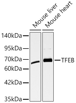
Western blot analysis of various lysates, using TFEB Rabbit pAb at 1:1000 dilution. Secondary antibody: HRP-conjugated Goat anti-Rabbit IgG (H+L) at 1:10000 dilution. Lysates/proteins: 25μg per lane. Blocking buffer: 3% nonfat dry milk in TBST. Detection: ECL Basic Kit. Exposure time: 60s.IHC
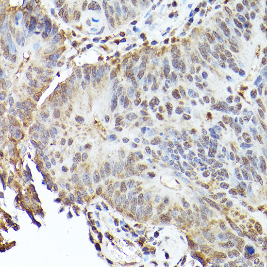
Immunohistochemistry analysis of paraffin embedded Human colon carcinoma using TFEB Rabbit pAb at dilution of 1:100 (40x lens). High pressure antigen retrieval performed with 0.01M Citrate buffer (pH 6.0) prior to IHC staining.IHC
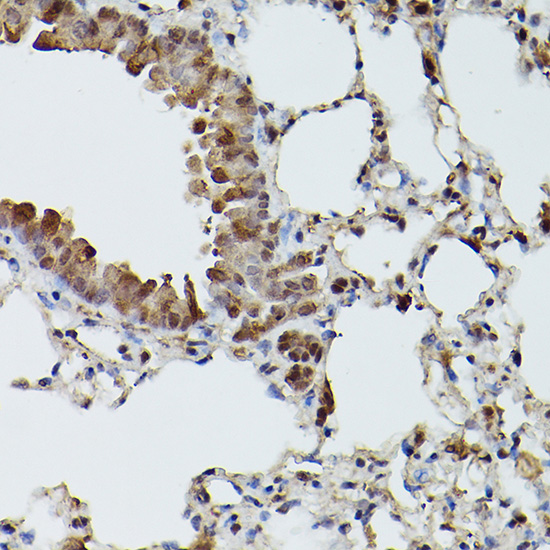
Immunohistochemistry analysis of paraffin embedded Mouse lung using TFEB Rabbit pAb at dilution of 1:100 (40x lens). High pressure antigen retrieval performed with 0.01M Citrate buffer (pH 6.0) prior to IHC staining.IHC
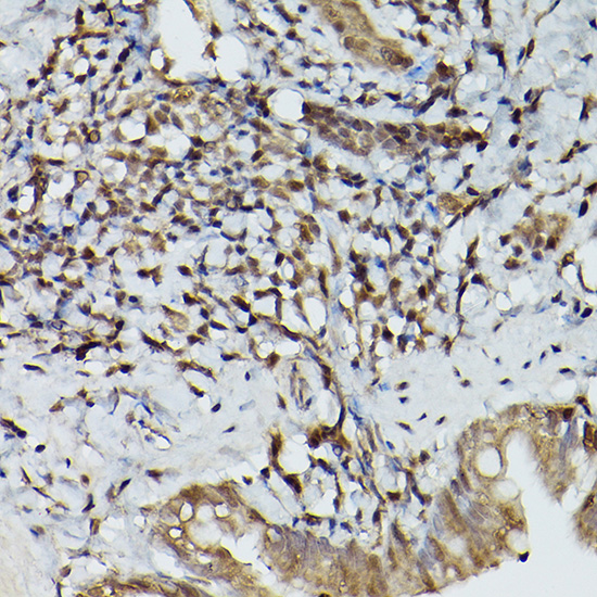
Immunohistochemistry analysis of paraffin embedded Rat lung using TFEB Rabbit pAb at dilution of 1:100 (40x lens). High pressure antigen retrieval performed with 0.01M Citrate buffer (pH 6.0) prior to IHC staining.ICC/IF
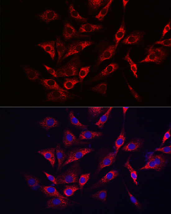
Immunofluorescence analysis of NIH/3T3 cells using TFEB Rabbit pAb at dilution of 1:100 (40x lens). Secondary antibody: Cy3-conjugated Goat anti-Rabbit IgG (H+L) at 1:500 dilution. Blue: DAPI for nuclear staining.| Product Name | TFEB Rabbit pAb |
|---|---|
| Antibody Type | Primary Antibodies |
| Immunogen | Recombinant protein. |
| Clonality | Polyclonal |
|---|---|
| Isotype | IgG |
| Host Species | Rabbit |
| Tested Applications | ICC/IFIHCWB |
| WB:1:500-1:1000 IHC:1:50-1:200 ICC/IF:1:50-1:200 |
|
| Species Reactivity | HumanMouseRat |
| Concentration | 1mg/ml |
| Purification | Affinity purified |
| Gene Symbol | TFEB |
|---|---|
| Gene Synonyms | TCFEB BHLHE35 ALPHATFEB |
| Gene Full Name | transcription factor EB |
| Gene Summary | Enables DNA-binding transcription factor activity; enzyme binding activity; and transcription cis-regulatory region binding activity. Involved in several processes, including antibacterial innate immune response; cellular response to amino acid starvation; and positive regulation of metabolic process. Located in cytosol and lysosomal membrane. Is active in nucleus. [provided by Alliance of Genome Resources, Jul 2025] |
| Molecular Weight(MW) | 52kDa(Observed MW 65-70kDa) |
| Cellular Localization | cytoplasm, cytosol, extracellular exosome, microtubule cytoskeleton, mitochondrial inner membrane, mitochondrial intermembrane space, mitochondrial matrix, mitochondrial nucleoid, mitochondrion, nucleus. |
WB
Western blot analysis of various lysates, using TFEB Rabbit pAb at 1:1000 dilution. Secondary antibody: HRP-conjugated Goat anti-Rabbit IgG (H+L) at 1:10000 dilution. Lysates/proteins: 25μg per lane. Blocking buffer: 3% nonfat dry milk in TBST. Detection: ECL Basic Kit. Exposure time: 60s.
IHC
Immunohistochemistry analysis of paraffin embedded Human colon carcinoma using TFEB Rabbit pAb at dilution of 1:100 (40x lens). High pressure antigen retrieval performed with 0.01M Citrate buffer (pH 6.0) prior to IHC staining.
IHC
Immunohistochemistry analysis of paraffin embedded Mouse lung using TFEB Rabbit pAb at dilution of 1:100 (40x lens). High pressure antigen retrieval performed with 0.01M Citrate buffer (pH 6.0) prior to IHC staining.
IHC
Immunohistochemistry analysis of paraffin embedded Rat lung using TFEB Rabbit pAb at dilution of 1:100 (40x lens). High pressure antigen retrieval performed with 0.01M Citrate buffer (pH 6.0) prior to IHC staining.
ICC/IF
Immunofluorescence analysis of NIH/3T3 cells using TFEB Rabbit pAb at dilution of 1:100 (40x lens). Secondary antibody: Cy3-conjugated Goat anti-Rabbit IgG (H+L) at 1:500 dilution. Blue: DAPI for nuclear staining.| Application Notes | WB:1:500-1:1000 IHC:1:50-1:200 ICC/IF:1:50-1:200 |
|---|
| Form | Liquid |
|---|---|
| Storage Instructions | Store at -20℃. Avoid freeze / thaw cycles. |
| Storage Buffer | PBS with 0.09% Sodium azide, 0.05% BSA, 50% glycerol, pH7.3. |


