WB
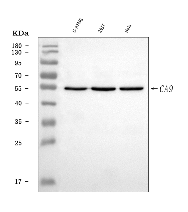
Western blot analysis of CA9 using anti-CA9 antibody. The sample well of each lane was loaded with 30 ug of sample under reducing conditions. Lane 1: U-87MG whole cell lysates, Lane 2: 293T whole cell lysates, Lane 3: Hela whole cell lysates. After electrophoresis, proteins were transferred to a membrane. Then the membrane was incubated with rabbit anti-CA9 antigen affinity purified polyclonal antibody at a dilution of 1:1000 and probed with a goat anti-rabbit IgG-HRP secondary antibody. The signal is developed using ECL Plus Western Blotting Substrate.IHC
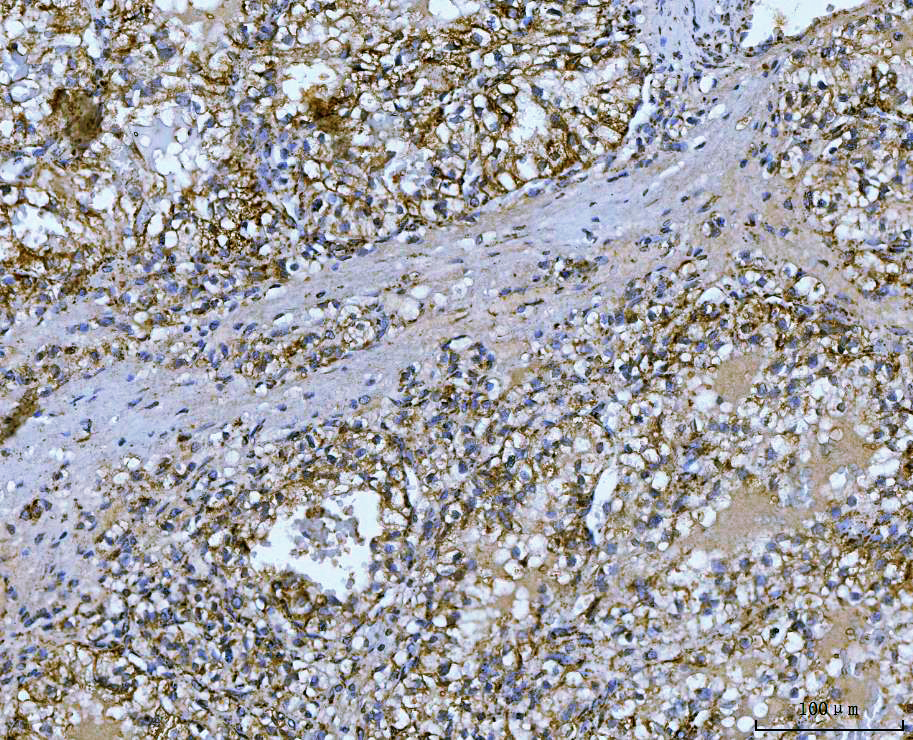
IHC analysis of CA9 using anti-CA9 antibody. CA9 was detected in a paraffin-embedded section of human renal adenocarcinoma tissue. Biotinylated goat anti-rabbit IgG was used as secondary antibody. The tissue section was incubated with rabbit anti-CA9 Antibody at a dilution of 1:200 and developed using Strepavidin-Biotin-Complex (SABC) with DAB as the chromogen.IHC
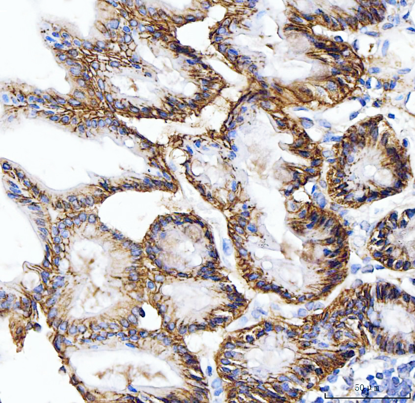
IHC analysis of CA9 using anti-CA9 antibody. CA9 was detected in a paraffin-embedded section of mouse stomach tissue. Biotinylated goat anti-rabbit IgG was used as secondary antibody. The tissue section was incubated with rabbit anti-CA9 Antibody at a dilution of 1:200 and developed using Strepavidin-Biotin-Complex (SABC) with DAB as the chromogen.FC
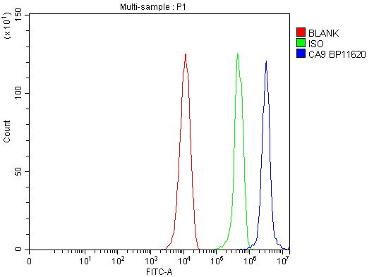
Flow Cytometry analysis of Raw264.7 cells using anti-CA9 antibody. Overlay histogram showing Raw264.7 cells stained with anti-CA9 antibody (Blue line). To facilitate intracellular staining, cells were fixed with 4% paraformaldehyde and permeabilized with permeabilization buffer. The cells were blocked with 10% normal goat serum. And then incubated with rabbit anti-CA9 Antibody at 1:100 dilution for 30 min at 20°C. DyLight®488 conjugated goat anti-rabbit IgG was used as secondary antibody at 1:100 dilution for 30 minutes at 20°C. Isotype control antibody (Green line) was rabbit IgG at 1:100 dilution used under the same conditions. Unlabelled sample without incubation with primary antibody and secondary antibody (Red line) was used as a blank control.| Product Name | Rabbit polyclonal antibody to CA9 |
|---|---|
| Antibody Type | Primary Antibodies |
| Immunogen | E.coli-derived human Carbonic Anhydrase 9/CA9 recombinant protein (Position: D146-D414). |
| Clonality | Polyclonal |
|---|---|
| Isotype | IgG |
| Host Species | Rabbit |
| Tested Applications | FCIHCWB |
| WB:1:500-1:2000 IHC:1:50-1:400 FC:1:50-1:200 |
|
| Species Reactivity | HumanMouse |
| Concentration | 0.5mg/ml |
| Purification | Affinity purified |
| Gene Symbol | CA9 |
|---|---|
| Gene Synonyms | MN, CAIX |
| Gene Full Name | carbonic anhydrase 9 |
| Gene Summary | Carbonic anhydrases (CAs) are a large family of zinc metalloenzymes that catalyze the reversible hydration of carbon dioxide. They participate in a variety of biological processes, including respiration, calcification, acid-base balance, bone resorption, and the formation of aqueous humor, cerebrospinal fluid, saliva, and gastric acid. They show extensive diversity in tissue distribution and in their subcellular localization. CA IX is a transmembrane protein and is one of only two tumor-associated carbonic anhydrase isoenzymes known. It is expressed in all clear-cell renal cell carcinoma, but is not detected in normal kidney or most other normal tissues. It may be involved in cell proliferation and transformation. This gene was mapped to 17q21.2 by fluorescence in situ hybridization, however, radiation hybrid mapping localized it to 9p13-p12. [provided by RefSeq, Jun 2014] |
| Alternative Names | CA IX CA9 CAIX Carbonate dehydratase IX Carbonic anhydrase 9 carbonic anhydrase IX G250 Membrane antigen MN MN P54/58N pMW1 RCC associated antigen G250 |
| Molecular Weight(MW) | 50 kDa(Observed MW 55 kDa) |
| Cellular Localization | Nucleus,Cell membrane,Cell projection, microvillus membrane. |
WB
Western blot analysis of CA9 using anti-CA9 antibody. The sample well of each lane was loaded with 30 ug of sample under reducing conditions. Lane 1: U-87MG whole cell lysates, Lane 2: 293T whole cell lysates, Lane 3: Hela whole cell lysates. After electrophoresis, proteins were transferred to a membrane. Then the membrane was incubated with rabbit anti-CA9 antigen affinity purified polyclonal antibody at a dilution of 1:1000 and probed with a goat anti-rabbit IgG-HRP secondary antibody. The signal is developed using ECL Plus Western Blotting Substrate.
IHC
IHC analysis of CA9 using anti-CA9 antibody. CA9 was detected in a paraffin-embedded section of human renal adenocarcinoma tissue. Biotinylated goat anti-rabbit IgG was used as secondary antibody. The tissue section was incubated with rabbit anti-CA9 Antibody at a dilution of 1:200 and developed using Strepavidin-Biotin-Complex (SABC) with DAB as the chromogen.
IHC
IHC analysis of CA9 using anti-CA9 antibody. CA9 was detected in a paraffin-embedded section of mouse stomach tissue. Biotinylated goat anti-rabbit IgG was used as secondary antibody. The tissue section was incubated with rabbit anti-CA9 Antibody at a dilution of 1:200 and developed using Strepavidin-Biotin-Complex (SABC) with DAB as the chromogen.
FC
Flow Cytometry analysis of Raw264.7 cells using anti-CA9 antibody. Overlay histogram showing Raw264.7 cells stained with anti-CA9 antibody (Blue line). To facilitate intracellular staining, cells were fixed with 4% paraformaldehyde and permeabilized with permeabilization buffer. The cells were blocked with 10% normal goat serum. And then incubated with rabbit anti-CA9 Antibody at 1:100 dilution for 30 min at 20°C. DyLight®488 conjugated goat anti-rabbit IgG was used as secondary antibody at 1:100 dilution for 30 minutes at 20°C. Isotype control antibody (Green line) was rabbit IgG at 1:100 dilution used under the same conditions. Unlabelled sample without incubation with primary antibody and secondary antibody (Red line) was used as a blank control.| Application Notes | WB:1:500-1:2000 IHC:1:50-1:400 FC:1:50-1:200 |
|---|
| Form | Liquid |
|---|---|
| Storage Instructions | 12 months from date of receipt, -20℃ as supplied. 6 months 2 to 8℃ after reconstitution. Avoid repeated freezing and thawing. |
| Storage Buffer | 500ug/ml antibody with PBS, 0.02% NaN3, 1 mg/ml BSA and 50% glycerol. |


