WB
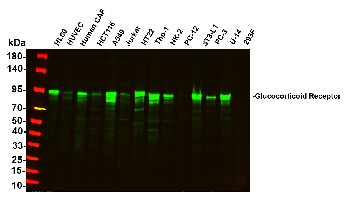
Various whole cell lysates were separated by 4-20% SDS-PAGE, and the primary antibody was used at 4°C, over night with a 1:2500 dilution . The Dylight 800-conjugated Goat anti-Rabbit antibody was used to detect the antibody. Lane1: HL60 - Human promyelocytic leukemia cell, Lane2: HUVEC - Human umbilical vein endothelial cell, Lane3: Human CAF - Human cancer-associated fibroblast, Lane4: HCT116 - Human colorectal carcinoma, Lane5: A549 - Human lung carcinoma, Lane6: Jurkat - Human T lymphocyte leukemia, Lane7: HT22 - Mouse hippocampal neuronal, Lane8: Thp-1 - Human monocytic leukemia, Lane9: HK-2 - Human proximal tubular epithelial, Lane10: PC-12 - Rat adrenal pheochromocytoma, Lane11: 3T3-L1 - Mouse embryonic fibroblast, Lane12: PC-3 - Human prostate adenocarcinoma, Lane13: U-14 - Mouse cervical carcinoma, Lane14: 293F - HEK293 derivative.WB
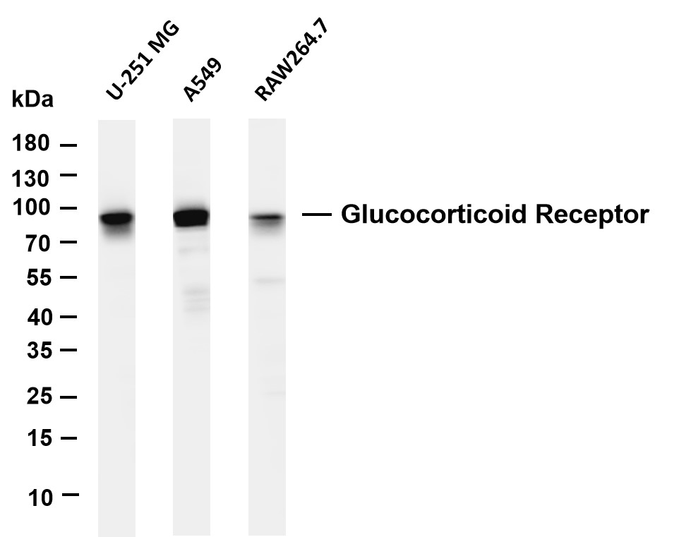
Various whole cell lysates were separated by 4-20% SDS-PAGE, and the membrane was blotted with anti-Glucocorticoid Receptor antibody. The HRP-conjugated Goat anti-Rabbit IgG(H + L) antibody was used to detect the antibody. Lane 1: U-251 MG, Lane 2: A549, Lane 3: RAW264.7.IHC
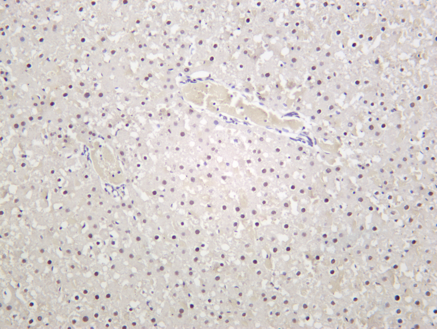
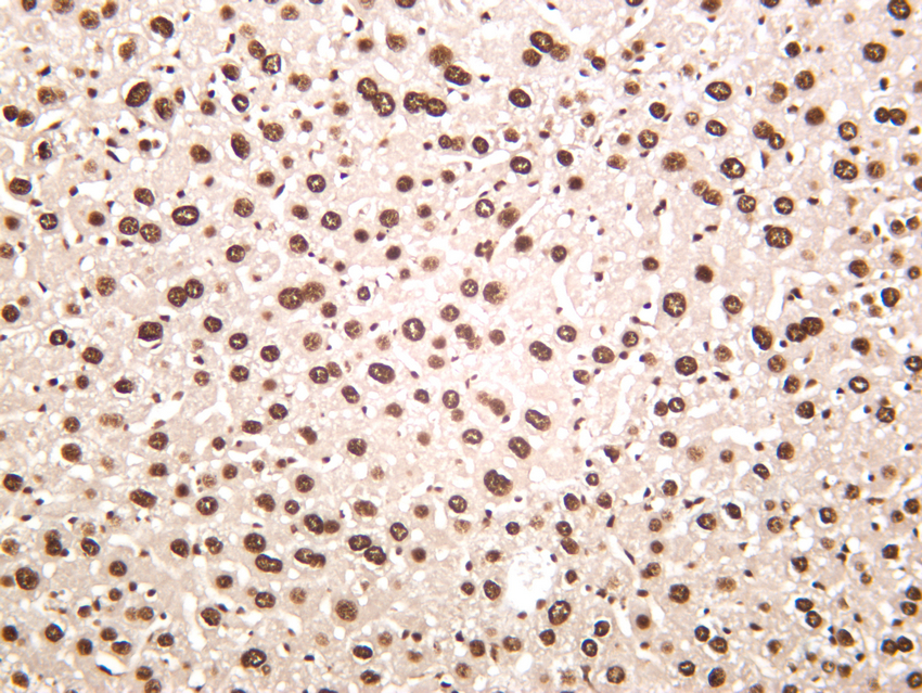
Rat liver was stained with anti-Glucocorticoid Receptor rabbit antibody.IHC
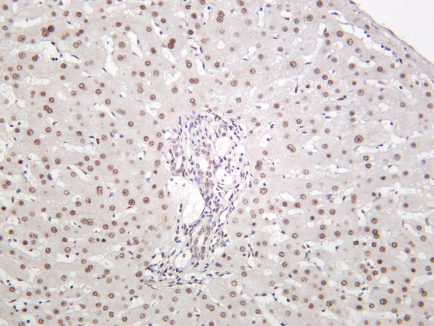
Human liver was stained with anti-Glucocorticoid Receptor rabbit antibody.IHC
Mouse liver was stained with anti-Glucocorticoid Receptor rabbit antibody.ICC/IF
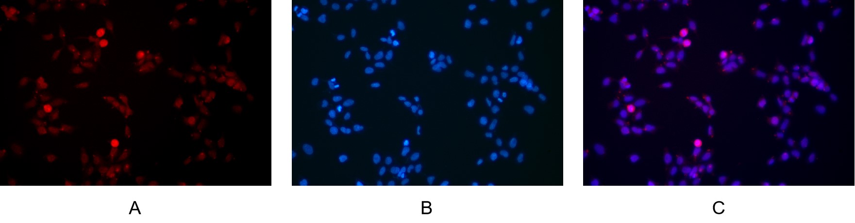
Immunofluorescence analysis of HEK293. Picture A:Glucocorticoid Receptor antibody (red). Picture B: DAPI (blue). Picture C: Merge of A+B.IF-P
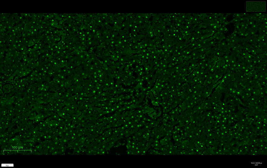
Rat liver was stained with anti-Glucocorticoid Receptor rabbit antibody| Product Name | Glucocorticoid Receptor Rabbit mAb |
|---|---|
| Antibody Type | Primary Antibodies |
| Clonality | monoclonal |
|---|---|
| Isotype | IgG |
| Host Species | Rabbit |
| Tested Applications | ICC/IFIF-PIHCWB |
| WB:1:1000-1:5000 IHC:1:1000-1:4000 ICC/IF:1:200-1:1000 IF-P:1:200-1:1000 |
|
| Species Reactivity | HumanMouseRat |
| Concentration | 1mg/ml |
| Purification | Protein A |
| Gene Symbol | NR3C1 |
|---|---|
| Gene Synonyms | GR GCR GRL GCCR GCRST |
| Gene Full Name | nuclear receptor subfamily 3 group C member 1 |
| Gene Summary | This gene encodes glucocorticoid receptor, which can function both as a transcription factor that binds to glucocorticoid response elements in the promoters of glucocorticoid responsive genes to activate their transcription, and as a regulator of other transcription factors. This receptor is typically found in the cytoplasm, but upon ligand binding, is transported into the nucleus. It is involved in inflammatory responses, cellular proliferation, and differentiation in target tissues. Mutations in this gene are associated with generalized glucocorticoid resistance. Alternative splicing of this gene results in transcript variants encoding either the same or different isoforms. Additional isoforms resulting from the use of alternate in-frame translation initiation sites have also been described, and shown to be functional, displaying diverse cytoplasm-to-nucleus trafficking patterns and distinct transcriptional activities (PMID:15866175). [provided by RefSeq, Feb 2011] |
| Alternative Names | NR3C1 GRL Glucocorticoid receptor GR Nuclear receptor subfamily 3 group C member 1 |
| Molecular Weight(MW) | 85kDa(Observed 94kDa) |
| Cellular Localization | Nucleus. |
WB
Various whole cell lysates were separated by 4-20% SDS-PAGE, and the primary antibody was used at 4°C, over night with a 1:2500 dilution . The Dylight 800-conjugated Goat anti-Rabbit antibody was used to detect the antibody. Lane1: HL60 - Human promyelocytic leukemia cell, Lane2: HUVEC - Human umbilical vein endothelial cell, Lane3: Human CAF - Human cancer-associated fibroblast, Lane4: HCT116 - Human colorectal carcinoma, Lane5: A549 - Human lung carcinoma, Lane6: Jurkat - Human T lymphocyte leukemia, Lane7: HT22 - Mouse hippocampal neuronal, Lane8: Thp-1 - Human monocytic leukemia, Lane9: HK-2 - Human proximal tubular epithelial, Lane10: PC-12 - Rat adrenal pheochromocytoma, Lane11: 3T3-L1 - Mouse embryonic fibroblast, Lane12: PC-3 - Human prostate adenocarcinoma, Lane13: U-14 - Mouse cervical carcinoma, Lane14: 293F - HEK293 derivative.
WB
Various whole cell lysates were separated by 4-20% SDS-PAGE, and the membrane was blotted with anti-Glucocorticoid Receptor antibody. The HRP-conjugated Goat anti-Rabbit IgG(H + L) antibody was used to detect the antibody. Lane 1: U-251 MG, Lane 2: A549, Lane 3: RAW264.7.
IHC
Rat liver was stained with anti-Glucocorticoid Receptor rabbit antibody.
IHC
Human liver was stained with anti-Glucocorticoid Receptor rabbit antibody.
IHC
Mouse liver was stained with anti-Glucocorticoid Receptor rabbit antibody.
ICC/IF
Immunofluorescence analysis of HEK293. Picture A:Glucocorticoid Receptor antibody (red). Picture B: DAPI (blue). Picture C: Merge of A+B.
IF-P
Rat liver was stained with anti-Glucocorticoid Receptor rabbit antibody| Application Notes | WB:1:1000-1:5000 IHC:1:1000-1:4000 ICC/IF:1:200-1:1000 IF-P:1:200-1:1000 |
|---|
| Form | Liquid |
|---|---|
| Storage Instructions | -15°C to -25°C/1 year(Do not lower than -25°C) |
| Storage Buffer | PBS, 50% glycerol, 0.05% Proclin 300, 0.05%BSA. |


