WB
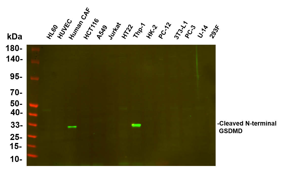
Various whole cell lysates were separated by 4-20% SDS-PAGE, and the primary antibody was used at 4°C, over night with a 1:5000 dilution . The Dylight 800-conjugated Goat anti-Rabbit antibody was used to detect the antibody. Lane1: HL60 - Human promyelocytic leukemia cell Lane2: HUVEC - Human umbilical vein endothelial cell Lane3: Human CAF - Human cancer-associated fibroblast Lane4: HCT116 - Human colorectal carcinoma Lane5: A549 - Human lung carcinoma Lane6: Jurkat - Human T lymphocyte leukemia Lane7: HT22 - Mouse hippocampal neuronal Lane8: Thp-1 - Human monocytic leukemia Lane9: HK-2 - Human proximal tubular epithelial Lane10: PC-12 - Rat adrenal pheochromocytoma Lane11: 3T3-L1 - Mouse embryonic fibroblast Lane12: PC-3 - Human prostate adenocarcinoma Lane13: U-14 - Mouse cervical carcinoma Lane14: 293F - HEK293 derivative.WB
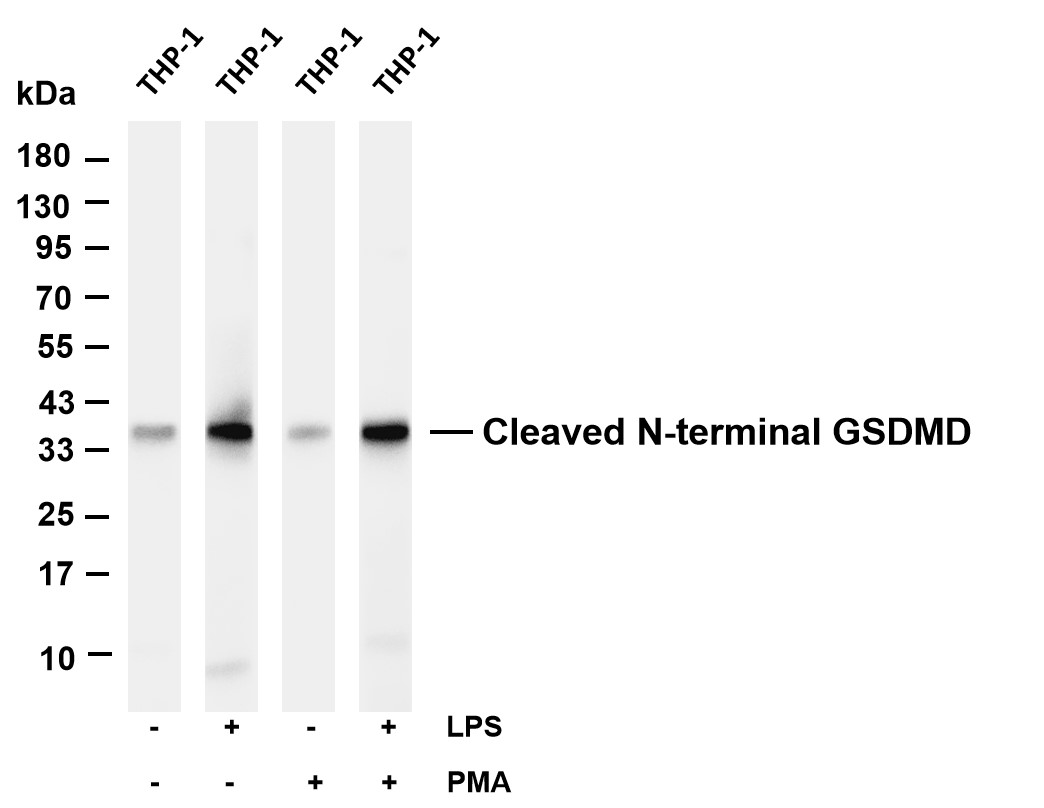
Various whole cell lysates were separated by 4-20% SDS-PAGE, and the membrane was blotted with anti-Cleaved N-terminal GSDMD antibody. The HRP-conjugated Goat anti-Rabbit IgG(H + L) antibody was used to detect the antibody. Lane 1: THP-1 Lane 2: THP-1 was treated with Lipopolysaccharides Lane 3: THP-1 was treated with Phorbol 12-myristate 13-acetate Lane 4: THP-1 was treated with Lipopolysaccharides and Phorbol 12-myristate 13-acetate.ICC/IF
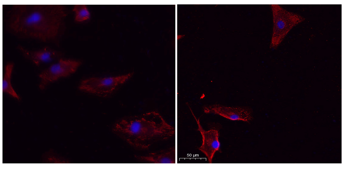
Immunofluorescence analysis of un-treated (left) A549 and UV treated(right) A549 cell. 1,primary Antibody was diluted at 1:200(4°C overnight). 2, Goat Anti Rabbit IgG (H&L) - Alexa Fluor 594 Secondary antibody was diluted at 1:1000(room temperature, 50min).3, Picture B: DAPI(blue) 10min.| Product Name | Cleaved N-terminal GSDMD Rabbit mAb |
|---|---|
| Antibody Type | Primary Antibodies |
| Clonality | monoclonal |
|---|---|
| Isotype | IgG |
| Host Species | Rabbit |
| Tested Applications | ICC/IFWB |
| WB:1:1000-1:5000 ICC/IF:1:200-1:1000 |
|
| Species Reactivity | Human |
| Concentration | 1mg/ml |
| Purification | Protein A |
| Gene Symbol | GSDMD |
|---|---|
| Gene Synonyms | DF5L DFNA5L FKSG10 GSDMDC1 |
| Gene Full Name | gasdermin D |
| Gene Summary | Gasdermin D is a member of the gasdermin family. Members of this family appear to play a role in regulation of epithelial proliferation. Gasdermin D has been suggested to act as a tumor suppressor. Alternatively spliced transcript variants have been described. [provided by RefSeq, Oct 2009] |
| Molecular Weight(MW) | 53kDa(Observed 35kDa) |
| Cellular Localization | Cytoplasm, cytosol. |
WB
Various whole cell lysates were separated by 4-20% SDS-PAGE, and the primary antibody was used at 4°C, over night with a 1:5000 dilution . The Dylight 800-conjugated Goat anti-Rabbit antibody was used to detect the antibody. Lane1: HL60 - Human promyelocytic leukemia cell Lane2: HUVEC - Human umbilical vein endothelial cell Lane3: Human CAF - Human cancer-associated fibroblast Lane4: HCT116 - Human colorectal carcinoma Lane5: A549 - Human lung carcinoma Lane6: Jurkat - Human T lymphocyte leukemia Lane7: HT22 - Mouse hippocampal neuronal Lane8: Thp-1 - Human monocytic leukemia Lane9: HK-2 - Human proximal tubular epithelial Lane10: PC-12 - Rat adrenal pheochromocytoma Lane11: 3T3-L1 - Mouse embryonic fibroblast Lane12: PC-3 - Human prostate adenocarcinoma Lane13: U-14 - Mouse cervical carcinoma Lane14: 293F - HEK293 derivative.
WB
Various whole cell lysates were separated by 4-20% SDS-PAGE, and the membrane was blotted with anti-Cleaved N-terminal GSDMD antibody. The HRP-conjugated Goat anti-Rabbit IgG(H + L) antibody was used to detect the antibody. Lane 1: THP-1 Lane 2: THP-1 was treated with Lipopolysaccharides Lane 3: THP-1 was treated with Phorbol 12-myristate 13-acetate Lane 4: THP-1 was treated with Lipopolysaccharides and Phorbol 12-myristate 13-acetate.
ICC/IF
Immunofluorescence analysis of un-treated (left) A549 and UV treated(right) A549 cell. 1,primary Antibody was diluted at 1:200(4°C overnight). 2, Goat Anti Rabbit IgG (H&L) - Alexa Fluor 594 Secondary antibody was diluted at 1:1000(room temperature, 50min).3, Picture B: DAPI(blue) 10min.| Application Notes | WB:1:1000-1:5000 ICC/IF:1:200-1:1000 |
|---|
| Form | Liquid |
|---|---|
| Storage Instructions | -15°C to -25°C/1 year(Do not lower than -25°C) |
| Storage Buffer | PBS, 50% glycerol, 0.05% Proclin 300, 0.05%BSA. |


