WB
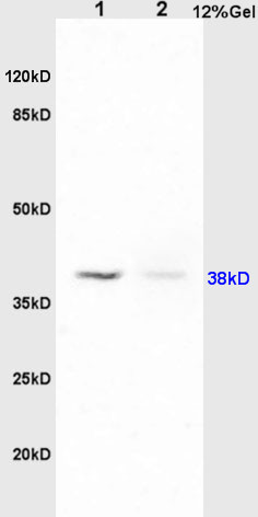
Sample: Lane1: Lung(Rat) Lysate at 30 ug, Lane2: Brain(Rat) Lysate at 30 ug, Primary: Anti-PTAFR at 1:200 dilution; Secondary: HRP conjugated Goat Anti-Rabbit IgG at 1: 3000 dilution.IHC
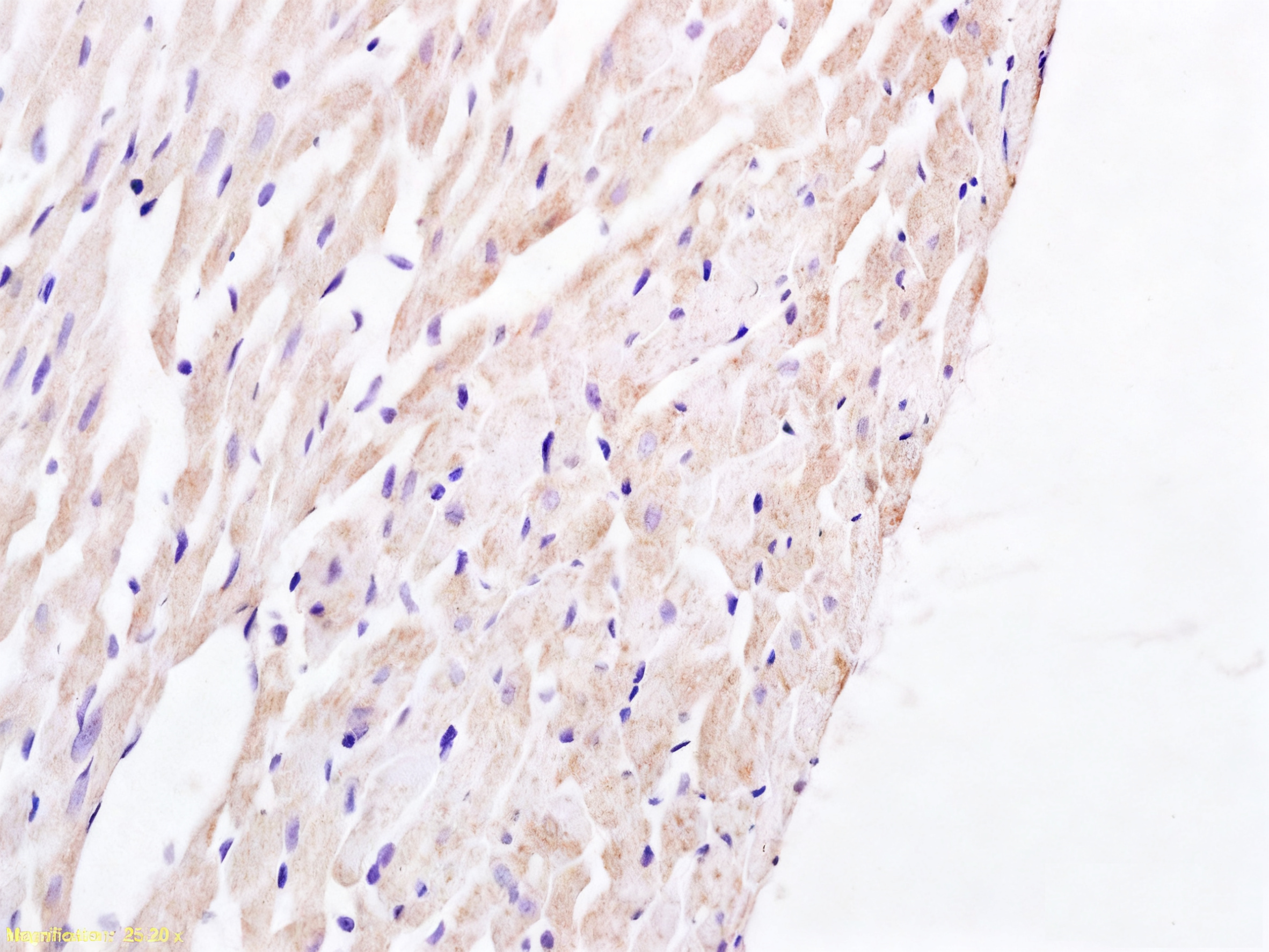
Tissue/cell: rat heart tissue; 4% Paraformaldehyde-fixed and paraffin-embedded; Antigen retrieval: citrate buffer ( 0.01M, pH 6.0 ), Boiling bathing for 15min; Block endogenous peroxidase by 3% Hydrogen peroxide for 30min; Blocking buffer (normal goat serum) at 37℃ for 20 min; Incubation: Anti-PTAFR Polyclonal Antibody 1:200, overnight at 4°C, followed by conjugation to the secondary antibody and DAB staining.FC
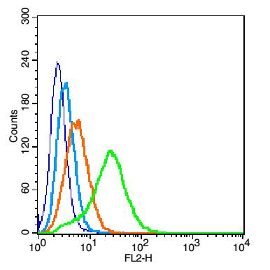
Blank control: 293T cells(blue). Primary Antibody:Rabbit Anti-PAFR/PAF antibody, Dilution: 1μg in 100 μL 1X PBS containing 0.5% BSA; Isotype Control Antibody: Rabbit IgG(orange) ,used under the same conditions ); Secondary Antibody: Goat anti-rabbit IgG-PE(white blue), Dilution: 1:200 in 1 X PBS containing 0.5% BSA. Protocol: The cells were fixed with 2% paraformaldehyde (10 min). Primary antibody (1μg /1x10^6 cells) were incubated for 30 min on the ice, followed by 1 X PBS containing 0.5% BSA + 1 0% goat serum (15 min) to block non-specific protein-protein interactions. Then the Goat Anti-rabbit IgG/PE antibody was added into the blocking buffer mentioned above to react with the primary antibody at 1/200 dilution for 30 min on ice. Acquisition of 20,000 events was performed.| Product Name | PAFR/PAF Receptor Rabbit pAb |
|---|---|
| Antibody Type | Primary Antibodies |
| Immunogen | KLH conjugated synthetic peptide derived from human PAFR : 231-342/342 |
| Clonality | Polyclonal |
|---|---|
| Isotype | IgG |
| Host Species | Rabbit |
| Tested Applications | FCIHCWB |
| WB:1:500-1:2000 IHC:1:100-1:500 FC:1ug/Test |
|
| Species Reactivity | HumanMouseRat |
| Concentration | 1mg/ml |
| Purification | Protein A |
| Gene Symbol | PTAFR |
|---|---|
| Gene Synonyms | PAFR |
| Gene Full Name | platelet activating factor receptor |
| Gene Summary | This gene encodes a seven-transmembrane G-protein-coupled receptor for platelet-activating factor (PAF) that localizes to lipid rafts and/or caveolae in the cell membrane. PAF (1-0-alkyl-2-acetyl-sn-glycero-3-phosphorylcholine) is a phospholipid that plays a significant role in oncogenic transformation, tumor growth, angiogenesis, metastasis, and pro-inflammatory processes. Binding of PAF to the PAF-receptor (PAFR) stimulates numerous signal transduction pathways including phospholipase C, D, A2, mitogen-activated protein kinases (MAPKs), and the phosphatidylinositol-calcium second messenger system. Following PAFR activation, cells become rapidly desensitized and this refractory state is dependent on PAFR phosphorylation, internalization, and down-regulation. Alternative splicing results in multiple transcript variants. [provided by RefSeq, Aug 2011] |
| Alternative Names | PTAFR_HUMAN PTAFR PAF-R PAFr PTAFR_MOUSE PTAFR_PIG PTAFR_RAT PTAFR_MACMU platelet activating factor receptor |
| Molecular Weight(MW) | 38kDa |
| Cellular Localization | Cell membrane. |
WB
Sample: Lane1: Lung(Rat) Lysate at 30 ug, Lane2: Brain(Rat) Lysate at 30 ug, Primary: Anti-PTAFR at 1:200 dilution; Secondary: HRP conjugated Goat Anti-Rabbit IgG at 1: 3000 dilution.
IHC
Tissue/cell: rat heart tissue; 4% Paraformaldehyde-fixed and paraffin-embedded; Antigen retrieval: citrate buffer ( 0.01M, pH 6.0 ), Boiling bathing for 15min; Block endogenous peroxidase by 3% Hydrogen peroxide for 30min; Blocking buffer (normal goat serum) at 37℃ for 20 min; Incubation: Anti-PTAFR Polyclonal Antibody 1:200, overnight at 4°C, followed by conjugation to the secondary antibody and DAB staining.
FC
Blank control: 293T cells(blue). Primary Antibody:Rabbit Anti-PAFR/PAF antibody, Dilution: 1μg in 100 μL 1X PBS containing 0.5% BSA; Isotype Control Antibody: Rabbit IgG(orange) ,used under the same conditions ); Secondary Antibody: Goat anti-rabbit IgG-PE(white blue), Dilution: 1:200 in 1 X PBS containing 0.5% BSA. Protocol: The cells were fixed with 2% paraformaldehyde (10 min). Primary antibody (1μg /1x10^6 cells) were incubated for 30 min on the ice, followed by 1 X PBS containing 0.5% BSA + 1 0% goat serum (15 min) to block non-specific protein-protein interactions. Then the Goat Anti-rabbit IgG/PE antibody was added into the blocking buffer mentioned above to react with the primary antibody at 1/200 dilution for 30 min on ice. Acquisition of 20,000 events was performed.| Application Notes | WB:1:500-1:2000 IHC:1:100-1:500 FC:1ug/Test |
|---|
| Form | Liquid |
|---|---|
| Storage Instructions | Shipped at 4℃. Store at -20℃ for one year. Avoid repeated freeze/thaw cycles. |
| Storage Buffer | 0.01M TBS (pH7.4) with 1% BSA, 0.02% Proclin300 and 50% Glycerol. |


