WB
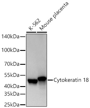
Western blot analysis of various lysates using Cytokeratin 18 Rabbit mAb at 1:12000 dilution incubated overnight at 4℃. Secondary antibody: HRP-conjugated Goat anti-Rabbit IgG (H+L) at 1:10000 dilution. Lysates/proteins: 25 μg per lane. Blocking buffer: 3% nonfat dry milk in TBST. Detection: ECL Basic Kit. Exposure time: 10sIHC
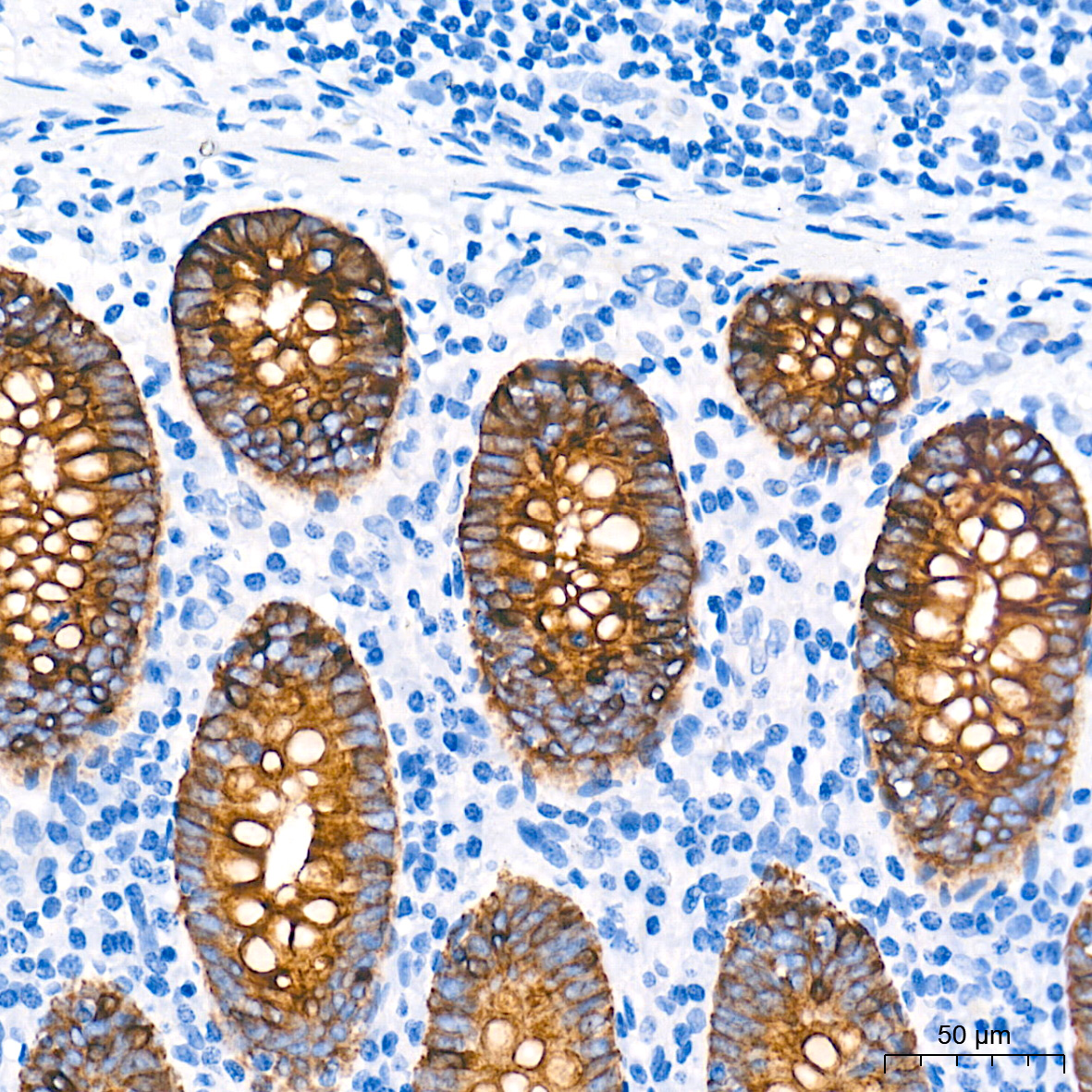
Immunohistochemistry analysis of paraffin embedded Human appendix tissue using Cytokeratin 18 Rabbit mAb at a dilution of 1:6000 (40x lens). High pressure antigen retrieval performed with 0.01M Tris EDTA Buffer (pH 9.0) prior to IHC staining.IHC
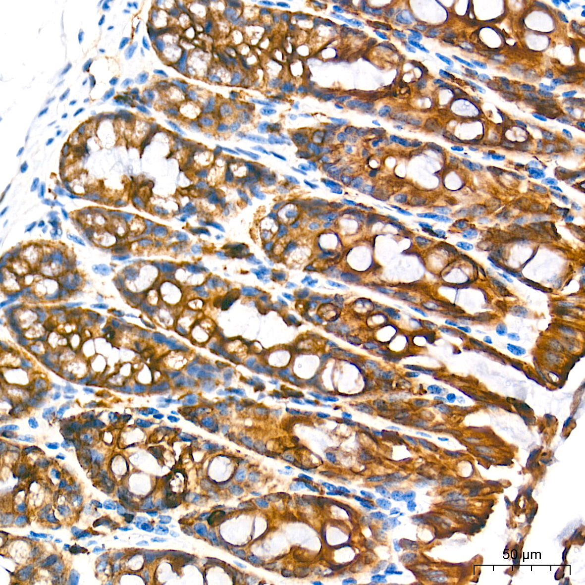
Immunohistochemistry analysis of paraffin embedded Mouse colon tissue using Cytokeratin 18 Rabbit mAb at a dilution of 1:6000 (40x lens). High pressure antigen retrieval performed with 0.01M Tris EDTA Buffer (pH 9.0) prior to IHC staining.IHC
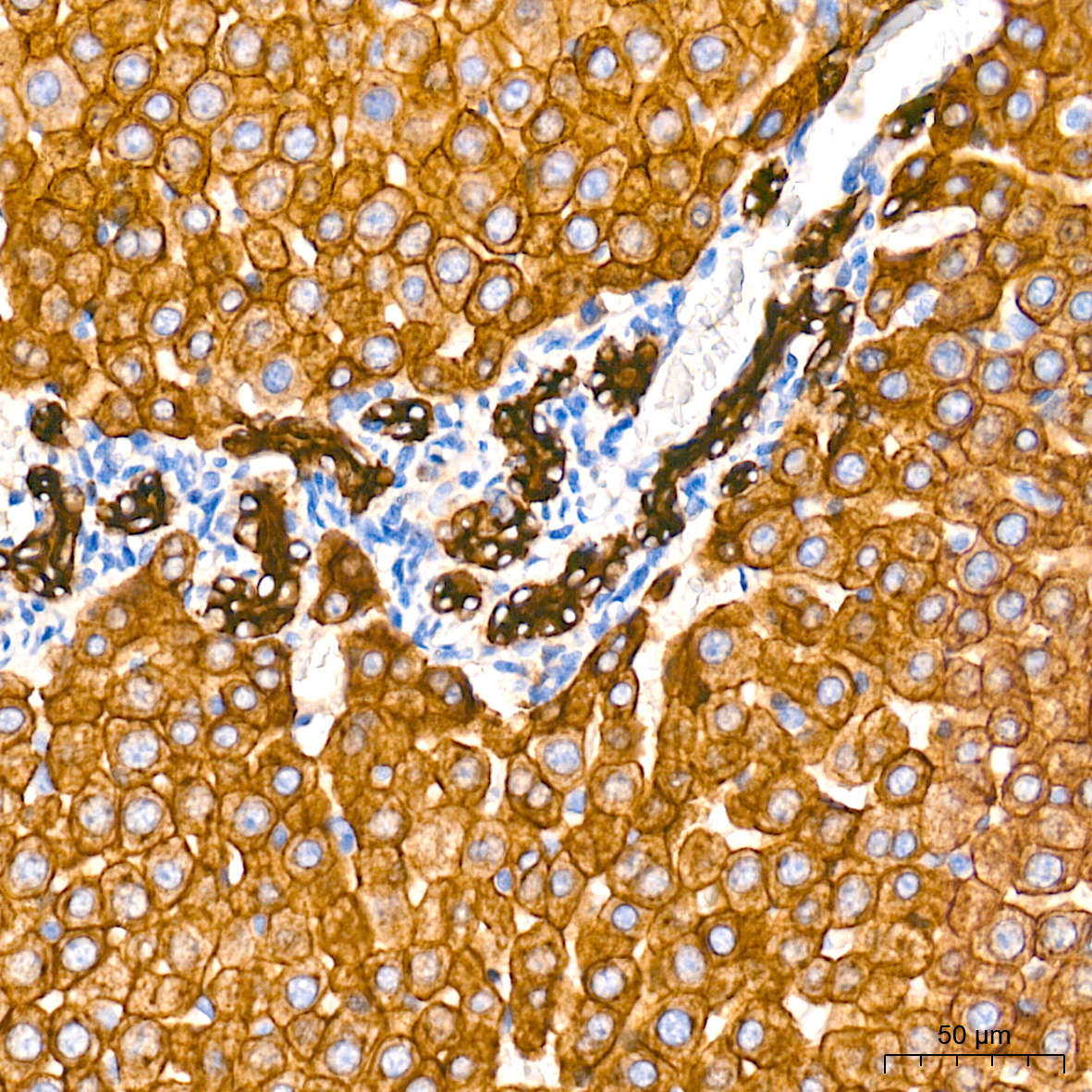
Immunohistochemistry analysis of paraffin embedded Rat liver tissue using Cytokeratin 18 Rabbit mAb at a dilution of 1:6000 (40x lens). High pressure antigen retrieval performed with 0.01M Tris-EDTA Buffer (pH 9.0) prior to IHC staining.ICC/IF
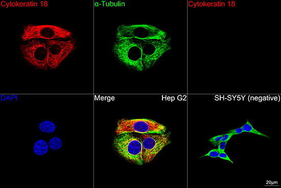
Confocal imaging of Hep G2 (positive) and SH-SY5Y (negative) cells using Cytokeratin 18 Rabbit mAb (dilution 1:800) followed by a further incubation with Cy3 Goat Anti-Rabbit IgG (H+L) (dilution 1:500) (Red). The cells were counterstained with α-Tubulin Mouse mAb (dilution 1:400) followed by incubation with Omnimabs® 488 Goat Anti-Mouse IgG(H&L) (dilution 1:500) (Green). DAPI was used for nuclear staining (Blue). Objective: 100x.IF-P
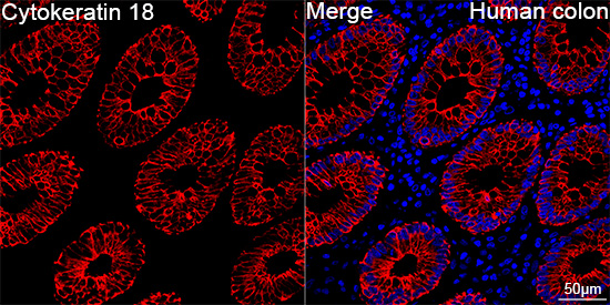
Confocal imaging of paraffin-embedded Human colon tissue using Cytokeratin 18 Rabbit mAb (dilution 1:800) followed by a further incubation with Cy3 Goat Anti-Rabbit IgG (H+L) (dilution 1:500) (Red). DAPI was used for nuclear staining (Blue). High pressure antigen retrieval performed with 0.01M Citrate Buffer (pH 6.0) prior to IF staining. Objective: 40x.IF-P
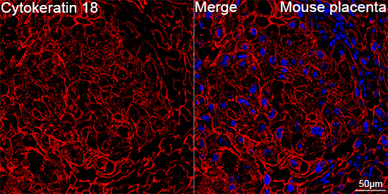
Confocal imaging of paraffin-embedded Mouse placenta tissue using Cytokeratin 18 Rabbit mAb (dilution 1:200) followed by a further incubation with Cy3 Goat Anti-Rabbit IgG (H+L) (dilution 1:500) (Red). DAPI was used for nuclear staining (Blue). High pressure antigen retrieval performed with 0.01M Citrate Buffer (pH 6.0) prior to IF staining. Objective: 40x.IF-P
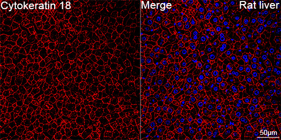
Confocal imaging of paraffin-embedded Rat liver tissue using Cytokeratin 18 Rabbit mAb (dilution 1:200) followed by a further incubation with Cy3 Goat Anti-Rabbit IgG (H+L) (dilution 1:500) (Red). DAPI was used for nuclear staining (Blue). High pressure antigen retrieval performed with 0.01M Citrate Buffer (pH 6.0) prior to IF staining. Objective: 40x.| Product Name | Cytokeratin 18 Rabbit mAb |
|---|---|
| Antibody Type | Primary Antibodies |
| Immunogen | Recombinant protein. |
| Clonality | Monoclonal |
|---|---|
| Isotype | IgG |
| Host Species | Rabbit |
| Tested Applications | ICC/IFIF-PIHCWB |
| WB:1:5000-1:30000 IHC:1:5000-1:30000 ICC/IF:1:200 - 1:2000 IF-P:1:200 - 1:2000 |
|
| Species Reactivity | HumanMouseRat |
| Concentration | 1mg/ml |
| Purification | Affinity purified |
| Gene Symbol | KRT18 |
|---|---|
| Gene Synonyms | K18 CK-18 CYK18 |
| Gene Full Name | keratin 18 |
| Gene Summary | KRT18 encodes the type I intermediate filament chain keratin 18. Keratin 18, together with its filament partner keratin 8, are perhaps the most commonly found members of the intermediate filament gene family. They are expressed in single layer epithelial tissues of the body. Mutations in this gene have been linked to cryptogenic cirrhosis. Two transcript variants encoding the same protein have been found for this gene. [provided by RefSeq, Jul 2008] |
| Alternative Names | K18 CK-18 CYK18 |
| Molecular Weight(MW) | 48kDa |
| Cellular Localization | adherens junction, cytoplasm, cytoskeleton, cytosol, extracellular exosome, microtubule organizing center, nuclear matrix, nucleolus, perinuclear region of cytoplasm, Cytoplasm, Nucleus, perinuclear region. |
WB
Western blot analysis of various lysates using Cytokeratin 18 Rabbit mAb at 1:12000 dilution incubated overnight at 4℃. Secondary antibody: HRP-conjugated Goat anti-Rabbit IgG (H+L) at 1:10000 dilution. Lysates/proteins: 25 μg per lane. Blocking buffer: 3% nonfat dry milk in TBST. Detection: ECL Basic Kit. Exposure time: 10s
IHC
Immunohistochemistry analysis of paraffin embedded Human appendix tissue using Cytokeratin 18 Rabbit mAb at a dilution of 1:6000 (40x lens). High pressure antigen retrieval performed with 0.01M Tris EDTA Buffer (pH 9.0) prior to IHC staining.
IHC
Immunohistochemistry analysis of paraffin embedded Mouse colon tissue using Cytokeratin 18 Rabbit mAb at a dilution of 1:6000 (40x lens). High pressure antigen retrieval performed with 0.01M Tris EDTA Buffer (pH 9.0) prior to IHC staining.
IHC
Immunohistochemistry analysis of paraffin embedded Rat liver tissue using Cytokeratin 18 Rabbit mAb at a dilution of 1:6000 (40x lens). High pressure antigen retrieval performed with 0.01M Tris-EDTA Buffer (pH 9.0) prior to IHC staining.
ICC/IF
Confocal imaging of Hep G2 (positive) and SH-SY5Y (negative) cells using Cytokeratin 18 Rabbit mAb (dilution 1:800) followed by a further incubation with Cy3 Goat Anti-Rabbit IgG (H+L) (dilution 1:500) (Red). The cells were counterstained with α-Tubulin Mouse mAb (dilution 1:400) followed by incubation with Omnimabs® 488 Goat Anti-Mouse IgG(H&L) (dilution 1:500) (Green). DAPI was used for nuclear staining (Blue). Objective: 100x.
IF-P
Confocal imaging of paraffin-embedded Human colon tissue using Cytokeratin 18 Rabbit mAb (dilution 1:800) followed by a further incubation with Cy3 Goat Anti-Rabbit IgG (H+L) (dilution 1:500) (Red). DAPI was used for nuclear staining (Blue). High pressure antigen retrieval performed with 0.01M Citrate Buffer (pH 6.0) prior to IF staining. Objective: 40x.
IF-P
Confocal imaging of paraffin-embedded Mouse placenta tissue using Cytokeratin 18 Rabbit mAb (dilution 1:200) followed by a further incubation with Cy3 Goat Anti-Rabbit IgG (H+L) (dilution 1:500) (Red). DAPI was used for nuclear staining (Blue). High pressure antigen retrieval performed with 0.01M Citrate Buffer (pH 6.0) prior to IF staining. Objective: 40x.
IF-P
Confocal imaging of paraffin-embedded Rat liver tissue using Cytokeratin 18 Rabbit mAb (dilution 1:200) followed by a further incubation with Cy3 Goat Anti-Rabbit IgG (H+L) (dilution 1:500) (Red). DAPI was used for nuclear staining (Blue). High pressure antigen retrieval performed with 0.01M Citrate Buffer (pH 6.0) prior to IF staining. Objective: 40x.| Application Notes | WB:1:5000-1:30000 IHC:1:5000-1:30000 ICC/IF:1:200 - 1:2000 IF-P:1:200 - 1:2000 |
|---|
| Form | Liquid |
|---|---|
| Storage Instructions | Store at -20℃. Avoid freeze / thaw cycles. |
| Storage Buffer | PBS containing 50% glycerol and 0.05% BSA, preserved with proclin300 or sodium azide (as specified on the Certificate of Analysis), pH 7.3. |


