WB
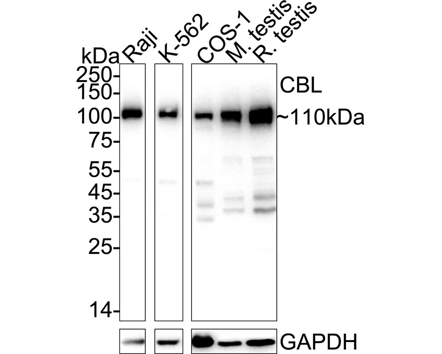
Western blot analysis of CBL on different lysates with Rabbit anti-CBL antibody at 1/5,000 dilution. Lane 1: Raji cell lysate (20 µg/Lane), Lane 2: K-562 cell lysate (20 µg/Lane), Lane 3: COS-1 cell lysate (20 µg/Lane), Lane 4: Mouse testis tissue lysate (30 µg/Lane), Lane 5: Rat testis tissue lysate (30 µg/Lane), Exposure time: 20 seconds; 4-20% SDS-PAGE gel. Proteins were transferred to a PVDF membrane and blocked with 5% NFDM/TBST for 1 hour at room temperature. The primary antibody at 1/5,000 dilution was used in primary antibody dilution at 4℃ overnight. Goat Anti-Rabbit IgG - HRP Secondary Antibody at 1/50,000 dilution was used for 1 hour at room temperature.ICC/IF
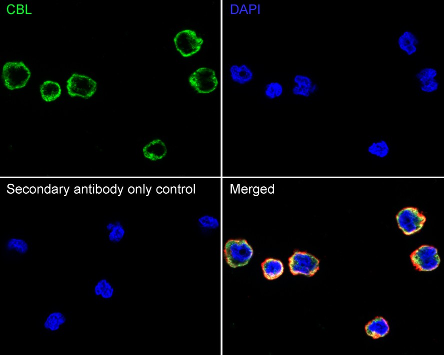
Immunocytochemistry analysis of Raji cells labeling CBL with Rabbit anti-CBL antibody at 1/100 dilution. Cells were fixed in 4% paraformaldehyde for 15 minutes at room temperature, permeabilized with 0.1% Triton X-100 in PBS for 15 minutes at room temperature, then blocked with 1% BSA in 10% negative goat serum for 1 hour at room temperature. Cells were then incubated with Rabbit anti-CBL antibody at 1/100 dilution in 1% BSA in PBST overnight at 4 ℃. Goat Anti-Rabbit IgG H&L (488) was used as the secondary antibody at 1/1,000 dilution. PBS instead of the primary antibody was used as the secondary antibody only control. Nuclear DNA was labelled in blue with DAPI. Beta tubulin (red) was stained at 1/100 dilution overnight at +4℃. Goat Anti-Mouse IgG H&L (594) was used as the secondary antibody at 1/1,000 dilution.FC
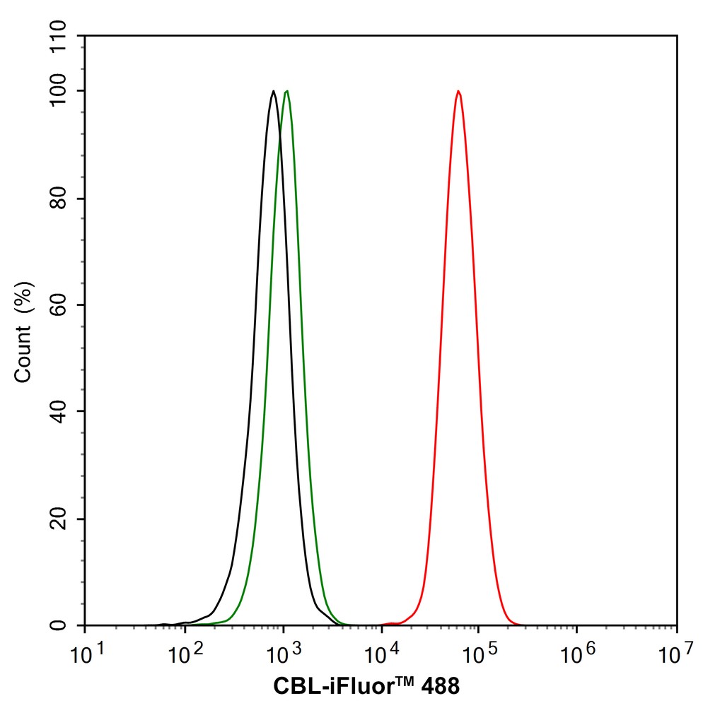
Flow cytometric analysis of Raji cells labeling CBL. Cells were fixed and permeabilized. Then stained with the primary antibody (1/1,000) (red) compared with Rabbit IgG Isotype Control (green). After incubation of the primary antibody at +4℃ for an hour, the cells were stained with a 488 conjugate-Goat anti-Rabbit IgG Secondary antibody at 1/1,000 dilution for 30 minutes at +4℃. Unlabelled sample was used as a control (cells without incubation with primary antibody; black).IP
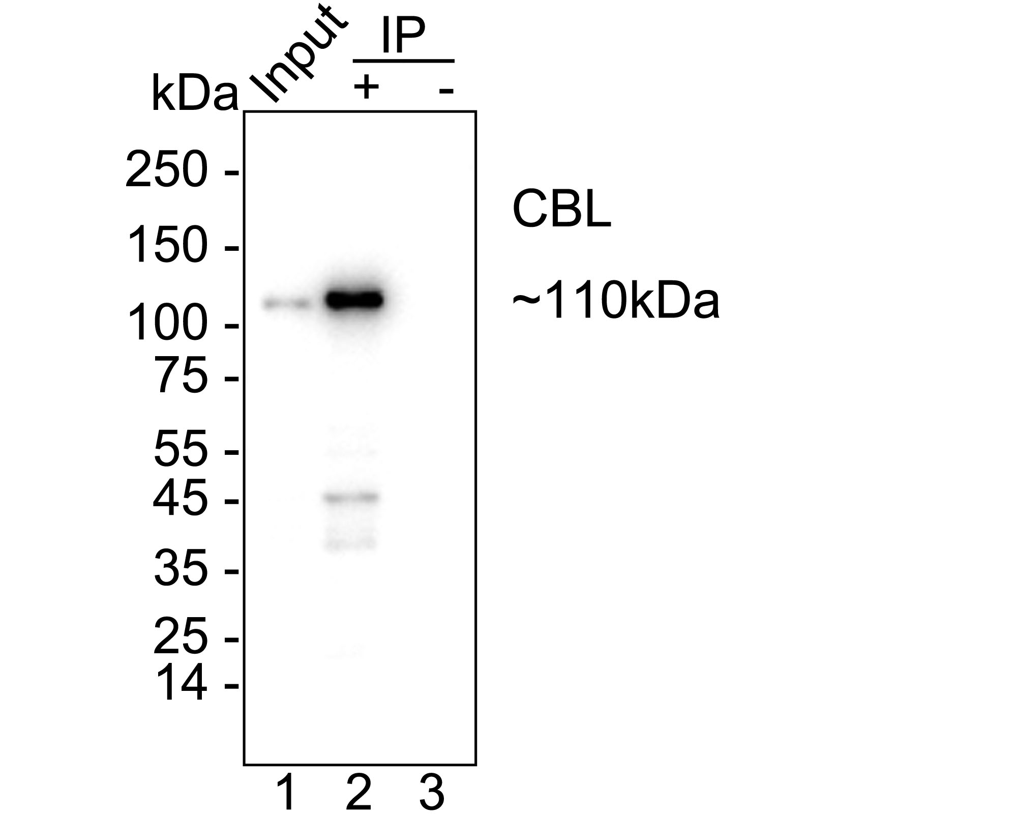
CBL was immunoprecipitated from 0.2 mg Raji cell lysate with Rabbit anti-CBL antibody at 2 µg/10 µl beads. Western blot was performed from the immunoprecipitate using Rabbit anti-CBL antibody at 1/5,000 dilution. HRP Conjugated Anti-Rabbit IgG for IP Nano-secondary antibody at 1/5,000 dilution was used for 1 hour at room temperature. Lane 1: Raji cell lysate (input), Lane 2: Rabbit anti-CBL antibody IP in Raji cell lysate, Lane 3: Rabbit IgG instead of Rabbit anti-CBL antibody in Raji cell lysate, Blocking/Dilution buffer: primary antibody dilution, Exposure time: 10 seconds.| Product Name | CBL Recombinant Rabbit Monoclonal Antibody |
|---|---|
| Antibody Type | Primary Antibodies |
| Immunogen | Recombinant protein within human CBL aa 441-840. |
| Clonality | Monoclonal |
|---|---|
| Isotype | IgG |
| Host Species | Rabbit |
| Tested Applications | FCICC/IFIPWB |
| WB:1:5000 ICC/IF:1:100 FC:1:1000 IP:1-2μg/sample |
|
| Species Reactivity | HumanMonkeyMouseRat |
| Concentration | 1mg/ml |
| Purification | Protein A |
| Gene Symbol | CBL |
|---|---|
| Gene Synonyms | CBL2 NSLL C-CBL RNF55 FRA11B |
| Gene Full Name | Cbl proto-oncogene |
| Gene Summary | This gene is a proto-oncogene that encodes a RING finger E3 ubiquitin ligase. The encoded protein is one of the enzymes required for targeting substrates for degradation by the proteasome. This protein mediates the transfer of ubiquitin from ubiquitin conjugating enzymes (E2) to specific substrates. This protein also contains an N-terminal phosphotyrosine binding domain that allows it to interact with numerous tyrosine-phosphorylated substrates and target them for proteasome degradation. As such it functions as a negative regulator of many signal transduction pathways. This gene has been found to be mutated or translocated in many cancers including acute myeloid leukaemia, and expansion of CGG repeats in the 5' UTR has been associated with Jacobsen syndrome. Mutations in this gene are also the cause of Noonan syndrome-like disorder. [provided by RefSeq, Jul 2016] |
| Alternative Names | 4732447J05Rik antibody C CBL antibody Cas Br M (murine) ecotropic retroviral transforming sequence antibody Casitas B lineage lymphoma proto oncogene antibody Casitas B-lineage lymphoma proto-oncogene antibody CBL 2 antibody cbl antibody CBL_HUMAN antibody CBL2 antibody E3 ubiquitin protein ligase CBL antibody 4732447J05Rik antibody C CBL antibody Cas Br M (murine) ecotropic retroviral transforming sequence antibody Casitas B lineage lymphoma proto oncogene antibody Casitas B-lineage lymphoma proto-oncogene antibody CBL 2 antibody cbl antibody CBL_HUMAN antibody CBL2 antibody E3 ubiquitin protein ligase CBL antibody E3 ubiquitin-protein ligase CBL antibody Oncogene CBL2 antibody Proto oncogene c CBL antibody Proto-oncogene c-CBL antibody RGD1561386 antibody RING finger protein 55 antibody RNF55 antibody Signal transduction protein CBL antibody |
| Molecular Weight(MW) | 100kDa(Observed band size: 110kDa) |
| Cellular Localization | Cytoplasm, Cell membrane, Cell projection, cilium, Golgi apparatus. |
WB
Western blot analysis of CBL on different lysates with Rabbit anti-CBL antibody at 1/5,000 dilution. Lane 1: Raji cell lysate (20 µg/Lane), Lane 2: K-562 cell lysate (20 µg/Lane), Lane 3: COS-1 cell lysate (20 µg/Lane), Lane 4: Mouse testis tissue lysate (30 µg/Lane), Lane 5: Rat testis tissue lysate (30 µg/Lane), Exposure time: 20 seconds; 4-20% SDS-PAGE gel. Proteins were transferred to a PVDF membrane and blocked with 5% NFDM/TBST for 1 hour at room temperature. The primary antibody at 1/5,000 dilution was used in primary antibody dilution at 4℃ overnight. Goat Anti-Rabbit IgG - HRP Secondary Antibody at 1/50,000 dilution was used for 1 hour at room temperature.
ICC/IF
Immunocytochemistry analysis of Raji cells labeling CBL with Rabbit anti-CBL antibody at 1/100 dilution. Cells were fixed in 4% paraformaldehyde for 15 minutes at room temperature, permeabilized with 0.1% Triton X-100 in PBS for 15 minutes at room temperature, then blocked with 1% BSA in 10% negative goat serum for 1 hour at room temperature. Cells were then incubated with Rabbit anti-CBL antibody at 1/100 dilution in 1% BSA in PBST overnight at 4 ℃. Goat Anti-Rabbit IgG H&L (488) was used as the secondary antibody at 1/1,000 dilution. PBS instead of the primary antibody was used as the secondary antibody only control. Nuclear DNA was labelled in blue with DAPI. Beta tubulin (red) was stained at 1/100 dilution overnight at +4℃. Goat Anti-Mouse IgG H&L (594) was used as the secondary antibody at 1/1,000 dilution.
FC
Flow cytometric analysis of Raji cells labeling CBL. Cells were fixed and permeabilized. Then stained with the primary antibody (1/1,000) (red) compared with Rabbit IgG Isotype Control (green). After incubation of the primary antibody at +4℃ for an hour, the cells were stained with a 488 conjugate-Goat anti-Rabbit IgG Secondary antibody at 1/1,000 dilution for 30 minutes at +4℃. Unlabelled sample was used as a control (cells without incubation with primary antibody; black).
IP
CBL was immunoprecipitated from 0.2 mg Raji cell lysate with Rabbit anti-CBL antibody at 2 µg/10 µl beads. Western blot was performed from the immunoprecipitate using Rabbit anti-CBL antibody at 1/5,000 dilution. HRP Conjugated Anti-Rabbit IgG for IP Nano-secondary antibody at 1/5,000 dilution was used for 1 hour at room temperature. Lane 1: Raji cell lysate (input), Lane 2: Rabbit anti-CBL antibody IP in Raji cell lysate, Lane 3: Rabbit IgG instead of Rabbit anti-CBL antibody in Raji cell lysate, Blocking/Dilution buffer: primary antibody dilution, Exposure time: 10 seconds.| Application Notes | WB:1:5000 ICC/IF:1:100 FC:1:1000 IP:1-2μg/sample |
|---|
| Form | Liquid |
|---|---|
| Storage Instructions | Store at +4℃ after thawing. Aliquot store at -20℃. Avoid repeated freeze / thaw cycles. |
| Storage Buffer | PBS (pH7.4), 0.1% BSA, 40% Glycerol. Preservative: 0.05% Sodium Azide. |


