WB
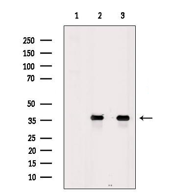
Western blot analysis of extracts from various samples, using Aquaporin 7 Antibody. Lane 1: EC304 cells, treated with blocking peptide; Lane 2: EC304 cells; Lane 3: P19 cells.IHC
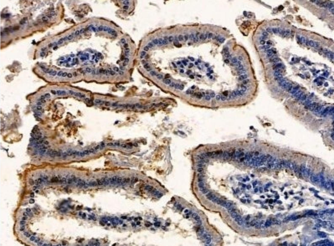
Aquaporin 7 Antibody at 1/100 staining mouse colorectal tissue by IHC-P. The sample was formaldehyde fixed and a heat mediated antigen retrieval step in citrate buffer was performed. The sample was then blocked and incubated with the primary antibody at 4°C overnight. An HRP conjugated anti-rabbit antibody was used as the secondary antibody.ICC/IF
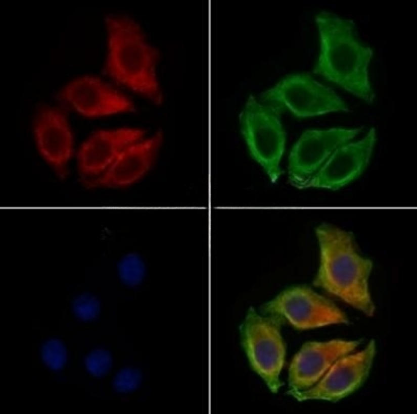
Aquaporin 7 Antibody staining HepG2 cells by IF/ICC. The samples were fixed with PFA and permeabilized in 0.1% Triton X-100,then blocked in 10% serum for 45 minutes at 25°C. Samples were then incubated with primary Ab(1:200) and mouse anti-beta tubulin Ab(1:200) for 1 hour at 37°C. An AlexaFluor594 conjugated goat anti-rabbit IgG(H+L) Ab(Red) and an AlexaFluor488 conjugated goat anti-mouse IgG(H+L) Ab(Green) were used as the secondary antibody. The nuclear counter stain is DAPI(blue).| Product Name | Rabbit polyclonal antibody to Aquaporin 7 |
|---|---|
| Antibody Type | Primary Antibodies |
| Immunogen | A synthesized peptide derived from human Aquaporin 7, corresponding to a region within N-terminal amino acids. |
| Clonality | Polyclonal |
|---|---|
| Isotype | IgG |
| Host Species | Rabbit |
| Tested Applications | ICC/IFIHCWB |
| WB:1:500-1:2000 IHC:1:50-1:200 ICC/IF:1:100-1:500 |
|
| Species Reactivity | HumanMouse |
| Concentration | 1mg/ml |
| Purification | Affinity purified |
| Gene Symbol | AQP7 |
|---|---|
| Gene Synonyms | AQP7L AQPap GLYCQTL |
| Gene Full Name | aquaporin 7 |
| Gene Summary | This gene encodes a member of the aquaporin family of water-selective membrane channels. The encoded protein localizes to the plasma membrane and allows movement of water, glycerol and urea across cell membranes. This gene is highly expressed in the adipose tissue where the encoded protein facilitates efflux of glycerol. In the proximal straight tubules of kidney, the encoded protein is localized to the apical membrane and prevents excretion of glycerol into urine. The encoded protein is present in spermatids, as well as in the testicular and epididymal spermatozoa suggesting an important role in late spermatogenesis. Alternative splicing of this gene results in multiple transcript variants encoding different isoforms. This gene is located adjacent to a related aquaporin gene on chromosome 9. Multiple pseudogenes of this gene have been identified. [provided by RefSeq, Dec 2015] |
| Alternative Names | AQP 7 AQP 7L AQP 9 AQP-7 Aqp7 AQP7_HUMAN AQP7L AQP9 AQPap Aquaglyceroporin-7 Aquaporin 7 like Aquaporin adipose Aquaporin-7 Aquaporin-7-like Aquaporin7 MGC149555 MGC149556 |
| Molecular Weight(MW) | 37kDa |
| Cellular Localization | Cell membrane>Multi-pass membrane protein. Cytoplasm>Cell cortex. Cytoplasmic vesicle membrane>Multi-pass membrane protein. Lipid droplet. |
WB
Western blot analysis of extracts from various samples, using Aquaporin 7 Antibody. Lane 1: EC304 cells, treated with blocking peptide; Lane 2: EC304 cells; Lane 3: P19 cells.
IHC
Aquaporin 7 Antibody at 1/100 staining mouse colorectal tissue by IHC-P. The sample was formaldehyde fixed and a heat mediated antigen retrieval step in citrate buffer was performed. The sample was then blocked and incubated with the primary antibody at 4°C overnight. An HRP conjugated anti-rabbit antibody was used as the secondary antibody.
ICC/IF
Aquaporin 7 Antibody staining HepG2 cells by IF/ICC. The samples were fixed with PFA and permeabilized in 0.1% Triton X-100,then blocked in 10% serum for 45 minutes at 25°C. Samples were then incubated with primary Ab(1:200) and mouse anti-beta tubulin Ab(1:200) for 1 hour at 37°C. An AlexaFluor594 conjugated goat anti-rabbit IgG(H+L) Ab(Red) and an AlexaFluor488 conjugated goat anti-mouse IgG(H+L) Ab(Green) were used as the secondary antibody. The nuclear counter stain is DAPI(blue).| Application Notes | WB:1:500-1:2000 IHC:1:50-1:200 ICC/IF:1:100-1:500 |
|---|
| Form | Liquid |
|---|---|
| Storage Instructions | Store at -20 °C. Stable for 12 months from date of receipt. |
| Storage Buffer | Rabbit IgG in phosphate buffered saline , pH 7.4, 150mM NaCl, 0.02% sodium azide and 50% glycerol. |


