WB
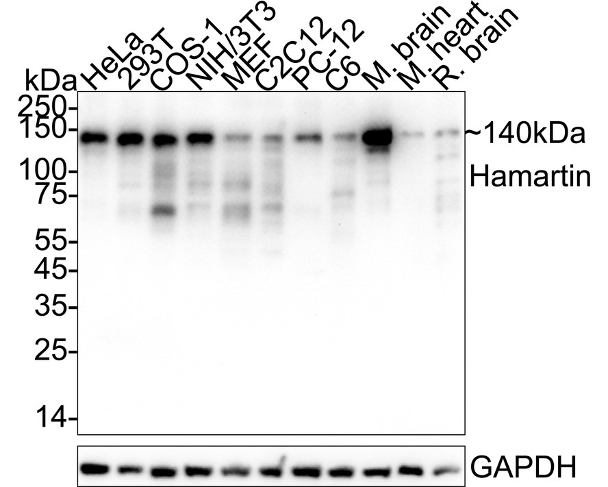
Western blot analysis of Hamartin on different lysates with Rabbit anti-Hamartin antibody at 1/20,000 dilution. Lane 1: HeLa cell lysate, Lane 2: 293T cell lysate, Lane 3: COS-1 cell lysate, Lane 4: NIH/3T3 cell lysate, Lane 5: MEF cell lysate, Lane 6: C2C12 cell lysate, Lane 7: PC-12 cell lysate, Lane 8: C6 cell lysate, Lane 9: Mouse brain tissue lysate, Lane 10: Mouse heart tissue lysate, Lane 11: Rat brain tissue lysate, Lysates/proteins at 20 µg/Lane. Exposure time: 1 minute; 4-20% SDS-PAGE gel. Proteins were transferred to a PVDF membrane and blocked with 5% NFDM/TBST for 1 hour at room temperature. The primary antibody at 1/20,000 dilution was used in primary antibody dilution at 4℃ overnight. Goat Anti-Rabbit IgG - HRP Secondary Antibody at 1/50,000 dilution was used for 1 hour at room temperature.IP
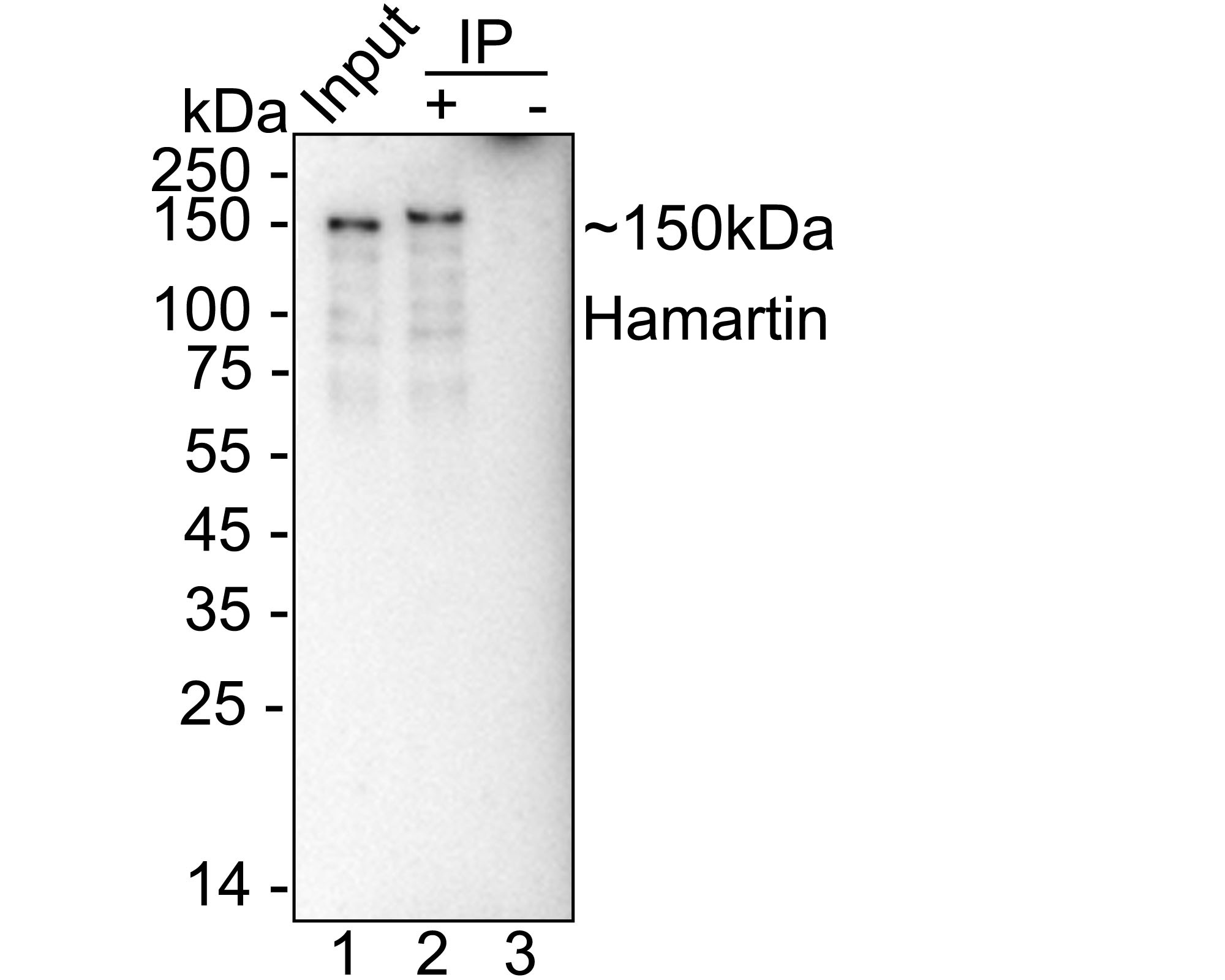
Hamartin was immunoprecipitated from 0.2 mg NIH/3T3 cell lysate with Rabbit anti-Hamartin antibody at 2 µg/10 µl beads. Western blot was performed from the immunoprecipitate using Rabbit anti-Hamartin antibody at 1/20,000 dilution. HRP Conjugated Anti-Rabbit IgG for IP Nano-secondary antibody at 1/5,000 dilution was used for 1 hour at room temperature. Lane 1: NIH/3T3 cell lysate (input), Lane 2: Rabbit anti-Hamartin antibody IP in NIH/3T3 cell lysate, Lane 3: Rabbit IgG instead of Rabbit anti-Hamartin antibody in NIH/3T3 cell lysate. Blocking/Dilution buffer: 5% NFDM/TBST Exposure time: 1 minute 50 seconds..| Product Name | Hamartin Recombinant Rabbit Monoclonal Antibody |
|---|---|
| Antibody Type | Primary Antibodies |
| Immunogen | Recombinant protein within mouse Hamartin aa 651-900. |
| Clonality | Monoclonal |
|---|---|
| Isotype | IgG |
| Host Species | Rabbit |
| Tested Applications | IPWB |
| WB:1:20000 IP:1-2μg/sample |
|
| Species Reactivity | HumanMonkeyMouseRat |
| Concentration | 1mg/ml |
| Purification | Protein A |
| Gene Symbol | TSC1 |
|---|---|
| Gene Full Name | TSC complex subunit 1 |
| Gene Summary | Enables ATPase inhibitor activity. Involved in associative learning; negative regulation of ATP-dependent activity; and protein stabilization. Acts upstream of or within several processes, including memory T cell differentiation; negative regulation of cell size; and nervous system development. Located in cytoplasm. Is active in postsynaptic density. Is expressed in several structures, including central nervous system; genitourinary system; hepatic primordium; liver; and retina. Used to study anterior segment dysgenesis; autism spectrum disorder; autosomal recessive polycystic kidney disease; spinal disease; and tuberous sclerosis. Human ortholog(s) of this gene implicated in intellectual disability; lymphangioleiomyomatosis; tuberous sclerosis; and tuberous sclerosis 1. Orthologous to human TSC1 (TSC complex subunit 1). [provided by Alliance of Genome Resources, Jul 2025] |
| Alternative Names | Hamartin antibody kiaa0243 antibody LAM antibody TSC antibody Tsc1 antibody Tsc1 gene antibody TSC1_HUMAN antibody Tuberous sclerosis 1 antibody Tuberous sclerosis 1 protein antibody tumor suppressor antibody |
| Molecular Weight(MW) | 130kDa(Observed band size: 140kDa) |
| Cellular Localization | Lysosome membrane, Cytoplasm, cytosol. |
WB
Western blot analysis of Hamartin on different lysates with Rabbit anti-Hamartin antibody at 1/20,000 dilution. Lane 1: HeLa cell lysate, Lane 2: 293T cell lysate, Lane 3: COS-1 cell lysate, Lane 4: NIH/3T3 cell lysate, Lane 5: MEF cell lysate, Lane 6: C2C12 cell lysate, Lane 7: PC-12 cell lysate, Lane 8: C6 cell lysate, Lane 9: Mouse brain tissue lysate, Lane 10: Mouse heart tissue lysate, Lane 11: Rat brain tissue lysate, Lysates/proteins at 20 µg/Lane. Exposure time: 1 minute; 4-20% SDS-PAGE gel. Proteins were transferred to a PVDF membrane and blocked with 5% NFDM/TBST for 1 hour at room temperature. The primary antibody at 1/20,000 dilution was used in primary antibody dilution at 4℃ overnight. Goat Anti-Rabbit IgG - HRP Secondary Antibody at 1/50,000 dilution was used for 1 hour at room temperature.
IP
Hamartin was immunoprecipitated from 0.2 mg NIH/3T3 cell lysate with Rabbit anti-Hamartin antibody at 2 µg/10 µl beads. Western blot was performed from the immunoprecipitate using Rabbit anti-Hamartin antibody at 1/20,000 dilution. HRP Conjugated Anti-Rabbit IgG for IP Nano-secondary antibody at 1/5,000 dilution was used for 1 hour at room temperature. Lane 1: NIH/3T3 cell lysate (input), Lane 2: Rabbit anti-Hamartin antibody IP in NIH/3T3 cell lysate, Lane 3: Rabbit IgG instead of Rabbit anti-Hamartin antibody in NIH/3T3 cell lysate. Blocking/Dilution buffer: 5% NFDM/TBST Exposure time: 1 minute 50 seconds..| Application Notes | WB:1:20000 IP:1-2μg/sample |
|---|
| Form | Liquid |
|---|---|
| Storage Instructions | Store at +4℃ after thawing. Aliquot store at -20℃. Avoid repeated freeze / thaw cycles. |
| Storage Buffer | PBS (pH7.4), 0.1% BSA, 40% Glycerol. Preservative: 0.05% Sodium Azide. |


