WB
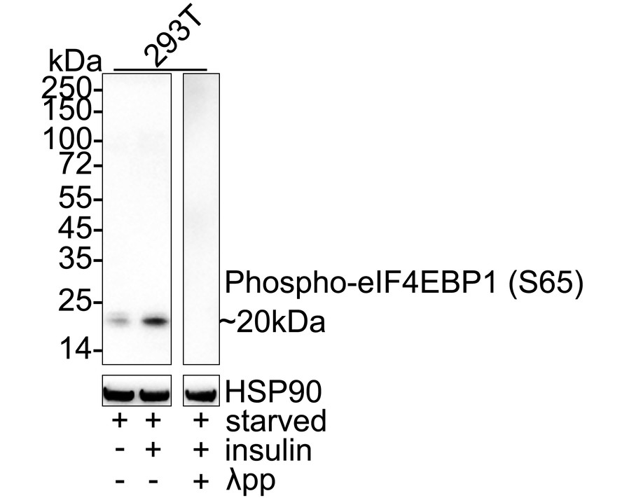
Western blot analysis of Phospho-eIF4EBP1 (S65) on different lysates with Rabbit anti-Phospho-eIF4EBP1 (S65) antibody at 1/1,000 dilution. Lane 1: 293T starved for 24 hours cell lysate, Lane 2: 293T starved for 24 hours then treated with 100nM insulin for 30 minutes cell lysate, Lane 3: 293T starved for 24 hours then treated with 100nM insulin for 30 minutes cell lysate, then the membrane treated with λpp for 1 hour, Lysates/proteins at 20 µg/Lane. Exposure time: 3 minutes; 4-20% SDS-PAGE gel. Proteins were transferred to a PVDF membrane and blocked with 5% NFDM/TBST for 1 hour at room temperature. The primary antibody at 1/1,000 dilution was used in primary antibody dilution (WB500D) at 4℃ overnight. Goat Anti-Rabbit IgG - HRP Secondary Antibody at 1/50,000 dilution was used for 1 hour at room temperature.ICC/IF
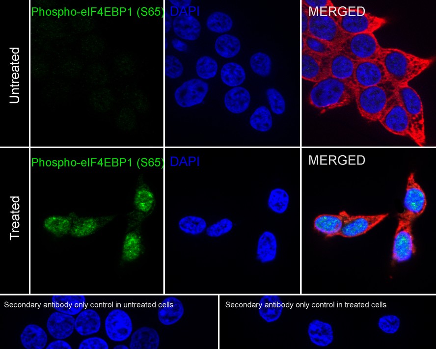
Immunocytochemistry analysis of 293T cells starved for 24 hours then treated with 100nM insulin for 30 minutes labeling Phospho-eIF4EBP1 (S65) with Rabbit anti-Phospho-eIF4EBP1 (S65) antibody at 1/1,000 dilution. Cells were fixed in 4% paraformaldehyde for 15 minutes at room temperature, permeabilized with 0.1% Triton X-100 in PBS for 15 minutes at room temperature, then blocked with 1% BSA in 10% negative goat serum for 1 hour at room temperature. Cells were then incubated with Rabbit anti-Phospho-eIF4EBP1 (S65) antibody at 1/1,000 dilution in 1% BSA in PBST overnight at 4 ℃. Goat Anti-Rabbit IgG H&L (488) was used as the secondary antibody at 1/1,000 dilution. PBS instead of the primary antibody was used as the secondary antibody only control. Nuclear DNA was labelled in blue with DAPI. Beta tubulin (red) was stained at 1/100 dilution overnight at +4℃. Goat Anti-Mouse IgG H&L (594) was used as the secondary antibody at 1/1,000 dilution.IP
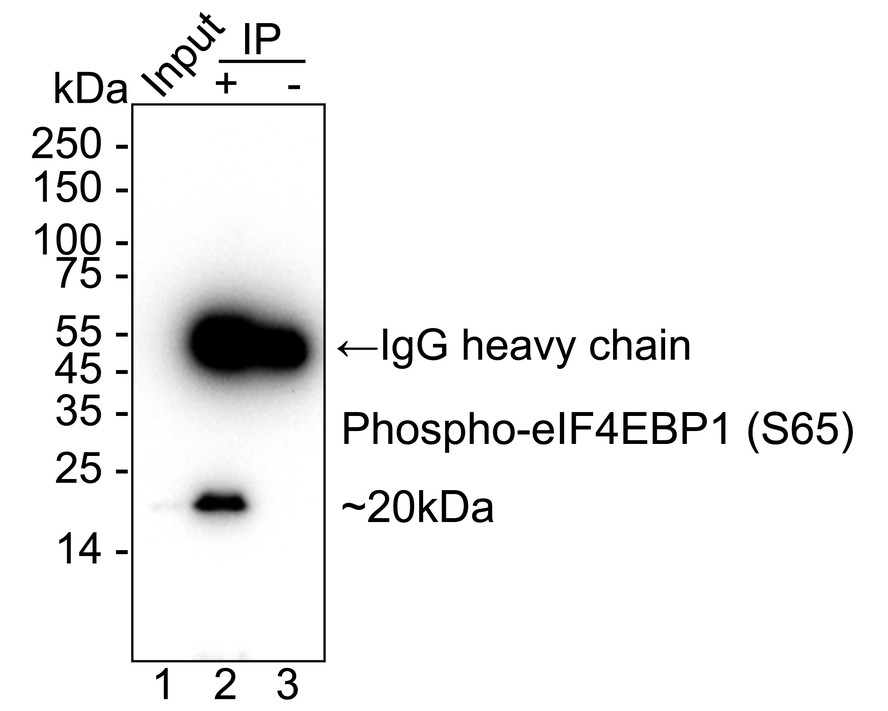
Phospho-eIF4EBP1 (S65) was immunoprecipitated from 0.2 mg 293T cell lysate with Rabbit anti-Phospho-eIF4EBP1 (S65) antibody at 2 µg/10 µl beads. Western blot was performed from the immunoprecipitate using Rabbit anti-Phospho-eIF4EBP1 (S65) antibody at 1/1,000 dilution. Alpaca anti-Rabbit IgG Fc secondary antibody at 1/50,000 dilution was used for 1 hour at room temperature. Lane 1: 293T cell lysate (input), Lane 2: Rabbit anti-Phospho-eIF4EBP1 (S65) antibody IP in 293T cell lysate, Lane 3: Rabbit IgG instead of Rabbit anti-Phospho-eIF4EBP1 (S65) antibody in 293T cell lysate, Blocking/Dilution buffer: 5% NFDM/TBST, Exposure time: 18 seconds.| Product Name | Phospho-eIF4EBP1 (S65) Recombinant Rabbit Monoclonal Antibody |
|---|---|
| Antibody Type | Primary Antibodies |
| Immunogen | Synthetic phospho-peptide corresponding to residues surrounding Ser65 of human eIF4EBP1. |
| Modification | p-S65 |
| Clonality | Monoclonal |
|---|---|
| Isotype | IgG |
| Host Species | Rabbit |
| Tested Applications | ICC/IFIPWB |
| WB:1:1000 ICC/IF:1:1000 IP:1-2μg/sample |
|
| Species Reactivity | Human |
| Concentration | 1mg/ml |
| Purification | Protein A |
| Gene Symbol | EIF4EBP1 |
|---|---|
| Gene Synonyms | BP-1 4EBP1 4E-BP1 PHAS-I |
| Gene Full Name | eukaryotic translation initiation factor 4E binding protein 1 |
| Gene Summary | This gene encodes one member of a family of translation repressor proteins. The protein directly interacts with eukaryotic translation initiation factor 4E (eIF4E), which is a limiting component of the multisubunit complex that recruits 40S ribosomal subunits to the 5' end of mRNAs. Interaction of this protein with eIF4E inhibits complex assembly and represses translation. This protein is phosphorylated in response to various signals including UV irradiation and insulin signaling, resulting in its dissociation from eIF4E and activation of mRNA translation. [provided by RefSeq, Jul 2008] |
| Alternative Names | 4E-BP1 antibody 4EBP1 antibody 4EBP1_HUMAN antibody BP 1 antibody eIF4E binding protein 1 antibody eIF4E-binding protein 1 antibody Eif4ebp1 antibody Eukaryotic translation initiation factor 4E-binding protein 1 antibody PHAS-I antibody PHASI antibody 4E-BP1 antibody 4EBP1 antibody 4EBP1_HUMAN antibody BP 1 antibody eIF4E binding protein 1 antibody eIF4E-binding protein 1 antibody Eif4ebp1 antibody Eukaryotic translation initiation factor 4E-binding protein 1 antibody PHAS-I antibody PHASI antibody Phosphorylated heat- and acid-stable protein regulated by insulin 1 antibody |
| Molecular Weight(MW) | 13 kDa(Observed band size: 20 kDa) |
| Cellular Localization | Cytoplasm, cytosol. |
WB
Western blot analysis of Phospho-eIF4EBP1 (S65) on different lysates with Rabbit anti-Phospho-eIF4EBP1 (S65) antibody at 1/1,000 dilution. Lane 1: 293T starved for 24 hours cell lysate, Lane 2: 293T starved for 24 hours then treated with 100nM insulin for 30 minutes cell lysate, Lane 3: 293T starved for 24 hours then treated with 100nM insulin for 30 minutes cell lysate, then the membrane treated with λpp for 1 hour, Lysates/proteins at 20 µg/Lane. Exposure time: 3 minutes; 4-20% SDS-PAGE gel. Proteins were transferred to a PVDF membrane and blocked with 5% NFDM/TBST for 1 hour at room temperature. The primary antibody at 1/1,000 dilution was used in primary antibody dilution (WB500D) at 4℃ overnight. Goat Anti-Rabbit IgG - HRP Secondary Antibody at 1/50,000 dilution was used for 1 hour at room temperature.
ICC/IF
Immunocytochemistry analysis of 293T cells starved for 24 hours then treated with 100nM insulin for 30 minutes labeling Phospho-eIF4EBP1 (S65) with Rabbit anti-Phospho-eIF4EBP1 (S65) antibody at 1/1,000 dilution. Cells were fixed in 4% paraformaldehyde for 15 minutes at room temperature, permeabilized with 0.1% Triton X-100 in PBS for 15 minutes at room temperature, then blocked with 1% BSA in 10% negative goat serum for 1 hour at room temperature. Cells were then incubated with Rabbit anti-Phospho-eIF4EBP1 (S65) antibody at 1/1,000 dilution in 1% BSA in PBST overnight at 4 ℃. Goat Anti-Rabbit IgG H&L (488) was used as the secondary antibody at 1/1,000 dilution. PBS instead of the primary antibody was used as the secondary antibody only control. Nuclear DNA was labelled in blue with DAPI. Beta tubulin (red) was stained at 1/100 dilution overnight at +4℃. Goat Anti-Mouse IgG H&L (594) was used as the secondary antibody at 1/1,000 dilution.
IP
Phospho-eIF4EBP1 (S65) was immunoprecipitated from 0.2 mg 293T cell lysate with Rabbit anti-Phospho-eIF4EBP1 (S65) antibody at 2 µg/10 µl beads. Western blot was performed from the immunoprecipitate using Rabbit anti-Phospho-eIF4EBP1 (S65) antibody at 1/1,000 dilution. Alpaca anti-Rabbit IgG Fc secondary antibody at 1/50,000 dilution was used for 1 hour at room temperature. Lane 1: 293T cell lysate (input), Lane 2: Rabbit anti-Phospho-eIF4EBP1 (S65) antibody IP in 293T cell lysate, Lane 3: Rabbit IgG instead of Rabbit anti-Phospho-eIF4EBP1 (S65) antibody in 293T cell lysate, Blocking/Dilution buffer: 5% NFDM/TBST, Exposure time: 18 seconds.| Application Notes | WB:1:1000 ICC/IF:1:1000 IP:1-2μg/sample |
|---|
| Form | Liquid |
|---|---|
| Storage Instructions | Store at +4℃ after thawing. Aliquot store at -20℃. Avoid repeated freeze / thaw cycles. |
| Storage Buffer | PBS (pH7.4), 0.1% BSA, 40% Glycerol. Preservative: 0.05% Sodium Azide. |


