WB
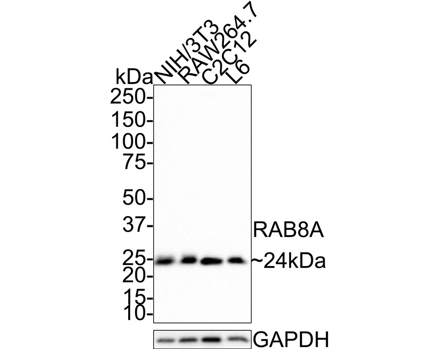
Western blot analysis of RAB8A on different lysates with Rabbit anti-RAB8A antibody at 1/500 dilution. Lane 1: NIH/3T3 cell lysate, Lane 2: RAW264.7 cell lysate, Lane 3: C2C12 cell lysate, Lane 4: L6 cell lysate, Lysates/proteins at 30 µg/Lane. Exposure time: 2 minutes 16 seconds; 4-20% SDS-PAGE gel. Proteins were transferred to a PVDF membrane and blocked with 5% NFDM/TBST for 1 hour at room temperature. The primary antibody at 1/500 dilution was used in 5% NFDM/TBST at room temperature for 2 hours. Goat Anti-Rabbit IgG - HRP Secondary Antibody at 1:100,000 dilution was used for 1 hour at room temperature.ICC/IF
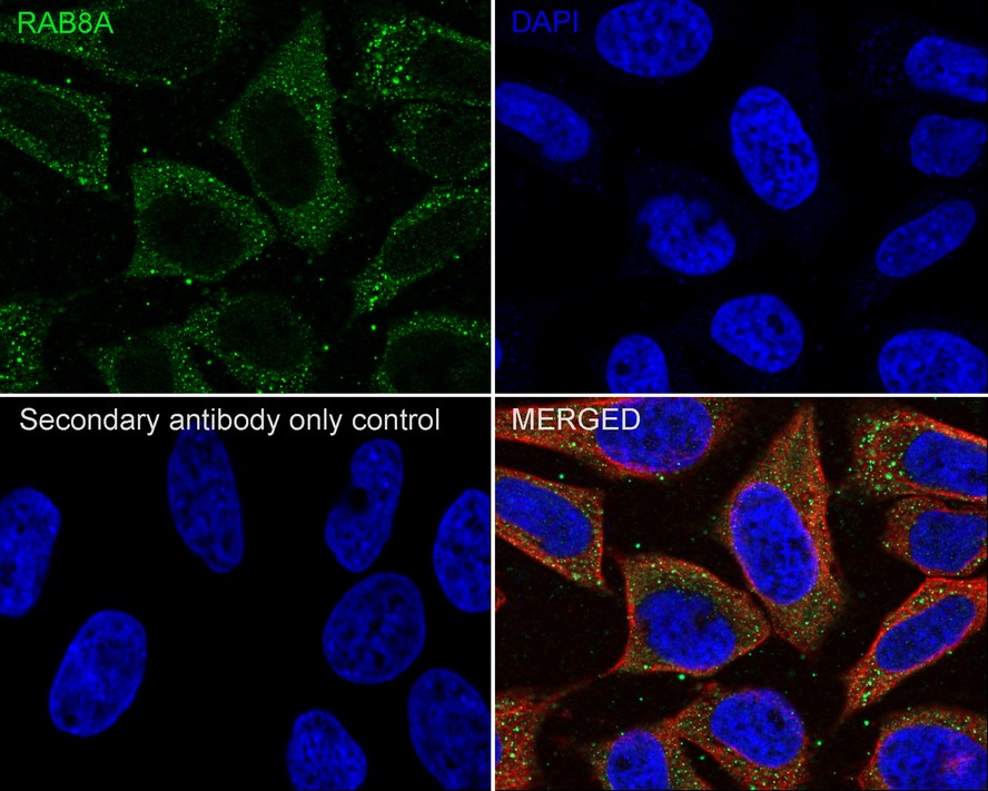
Immunocytochemistry analysis of HeLa cells labeling RAB8A with Rabbit anti-RAB8A antibody at 1/100 dilution. Cells were fixed in 4% paraformaldehyde for 10 minutes at 37 ℃, permeabilized with 0.05% Triton X-100 in PBS for 20 minutes, and then blocked with 2% negative goat serum for 30 minutes at room temperature. Cells were then incubated with Rabbit anti-RAB8A antibody at 1/100 dilution in 2% negative goat serum overnight at 4 ℃. Goat Anti-Rabbit IgG H&L (488) was used as the secondary antibody at 1/1,000 dilution. Nuclear DNA was labelled in blue with DAPI. Beta tubulin (red) was stained at 1/200 dilution overnight at +4℃. Goat Anti-Mouse IgG H&L (594) was used as the secondary antibody at 1/1,000 dilution.FC
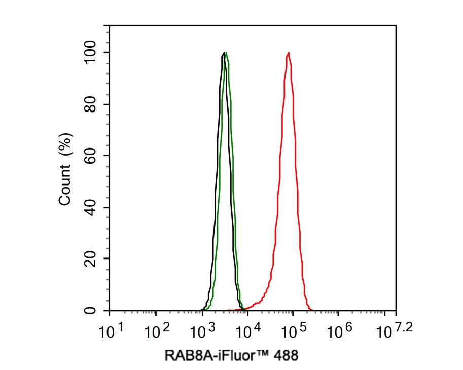
Flow cytometric analysis of HeLa cells labeling RAB8A. Cells were fixed and permeabilized. Then stained with the primary antibody (1ug/ml) (red) compared with Rabbit IgG Isotype Control (green). After incubation of the primary antibody at +4℃ for an hour, the cells were stained with a 488 conjugate-Goat anti-Rabbit IgG Secondary antibody at 1/1,000 dilution for 30 minutes at +4℃. Unlabelled sample was used as a control (cells without incubation with primary antibody; black).IP
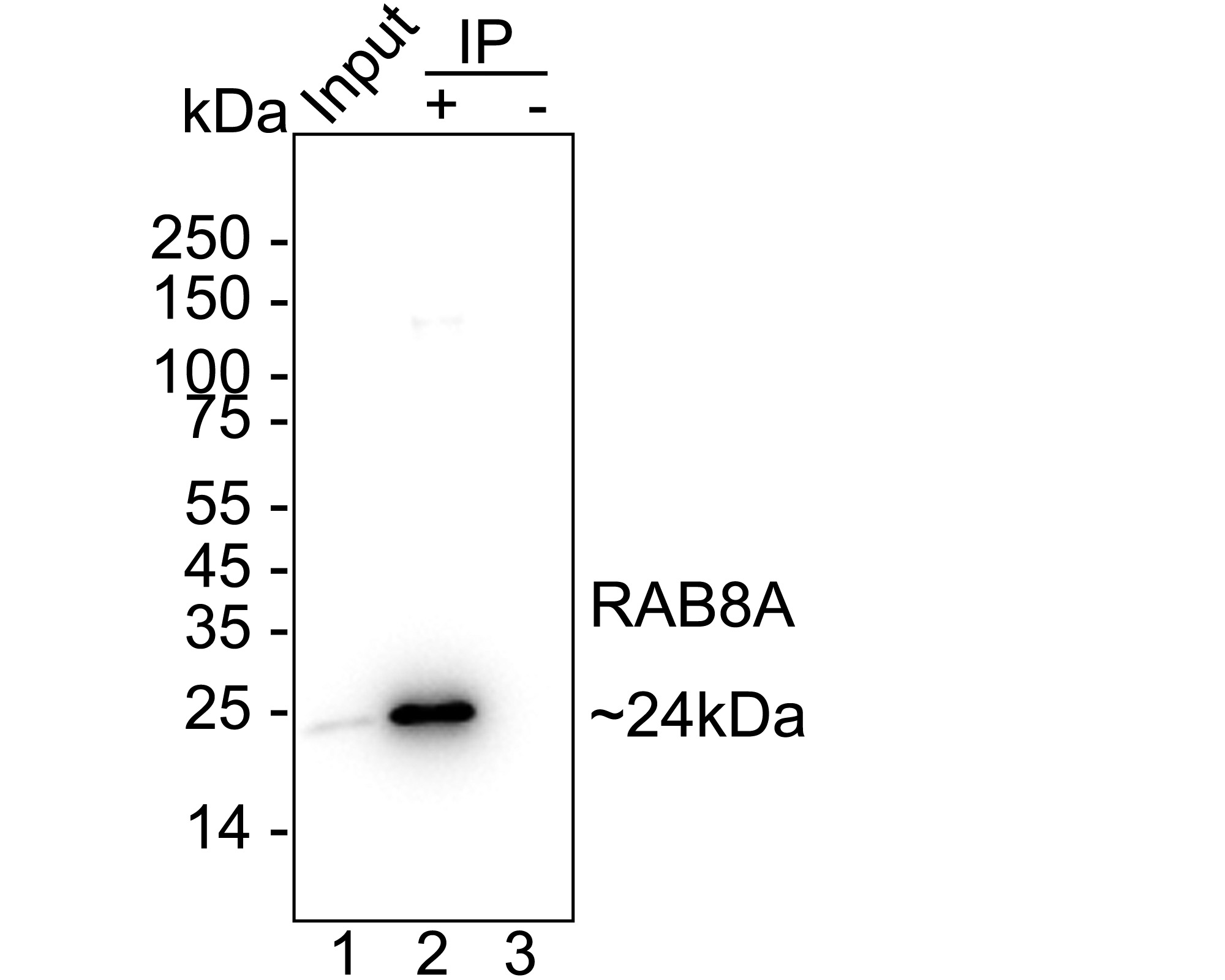
RAB8A was immunoprecipitated from 0.2 mg HeLa cell lysate with Rabbit anti-RAB8A antibody at 2 µg/10 µl beads. Western blot was performed from the immunoprecipitate using Rabbit anti-RAB8A antibody at 1/1,000 dilution. Anti-Rabbit IgG for IP Nano-secondary antibody at 1/5,000 dilution was used for 1 hour at room temperature. Lane 1: HeLa cell lysate (input), Lane 2: Rabbit anti-RAB8A antibody IP in HeLa cell lysate, Lane 3: Rabbit IgG instead of Rabbit anti-RAB8A antibody in HeLa cell lysate, Blocking/Dilution buffer: 5% NFDM/TBST, Exposure time: 4 seconds.| Product Name | RAB8A Recombinant Rabbit Monoclonal Antibody |
|---|---|
| Antibody Type | Primary Antibodies |
| Immunogen | Synthetic peptide within human RAB8A aa 151-200/207. |
| Clonality | monoclonal |
|---|---|
| Isotype | IgG |
| Host Species | Rabbit |
| Tested Applications | FCICC/IFIPWB |
| WB:1:1000 ICC/IF:1:100 FC:1:500-1:1000 IP:1-2μg/sample |
|
| Species Reactivity | HumanMouseRat |
| Concentration | 1mg/ml |
| Purification | Protein A |
| Gene Symbol | RAB8A |
|---|---|
| Gene Synonyms | MEL RAB8 |
| Gene Full Name | RAB8A, member RAS oncogene family |
| Gene Summary | The protein encoded by this gene is a member of the RAS superfamily which are small GTP/GDP-binding proteins with an average size of 200 amino acids. The RAS-related proteins of the RAB/YPT family may play a role in the transport of proteins from the endoplasmic reticulum to the Golgi and the plasma membrane. This protein shares 97%, 96%, and 51% similarity with the dog RAB8, mouse MEL, and mouse YPT1 proteins, respectively and contains the 4 GTP/GDP-binding sites that are present in all the RAS proteins. The putative effector-binding site of this protein is similar to that of the RAB/YPT proteins. However, this protein contains a C-terminal CAAX motif that is characteristic of many RAS superfamily members but which is not found in YPT1 and the majority of RAB proteins. Although this gene was isolated as a transforming gene from a melanoma cell line, no linkage between MEL and malignant melanoma has been demonstrable. This oncogene is located 800 kb distal to MY09B on chromosome 19p13.1. [provided by RefSeq, Jul 2008] |
| Alternative Names | AA409338 antibody MEL antibody Mel transforming oncogene (derived from cell line NK14) RAB8 homolog antibody Mel transforming oncogene (derived from cell line NK14) antibody Mel transforming oncogene (RAB8 homolog) antibody Mel transforming oncogene antibody MGC124948 antibody Oncogene c mel antibody Oncogene c-mel antibody RAB8 antibody AA409338 antibody MEL antibody Mel transforming oncogene (derived from cell line NK14) RAB8 homolog antibody Mel transforming oncogene (derived from cell line NK14) antibody Mel transforming oncogene (RAB8 homolog) antibody Mel transforming oncogene antibody MGC124948 antibody Oncogene c mel antibody Oncogene c-mel antibody RAB8 antibody RAB8A antibody RAB8A member RAS oncogene family antibody RAB8A_HUMAN antibody Ras associated protein RAB8 antibody Ras related protein Rab 8A antibody Ras-related protein Rab-8A antibody |
| Molecular Weight(MW) | 121kDa(Observed band size: 150 kDa) |
| Cellular Localization | Cell membrane, golgi apparatus, recycling endosome membrane, cytoplasm, centriole, cilium basal body, cilium axoneme, cilium, phagosome, phagosome membrane, midbody. |
WB
Western blot analysis of RAB8A on different lysates with Rabbit anti-RAB8A antibody at 1/500 dilution. Lane 1: NIH/3T3 cell lysate, Lane 2: RAW264.7 cell lysate, Lane 3: C2C12 cell lysate, Lane 4: L6 cell lysate, Lysates/proteins at 30 µg/Lane. Exposure time: 2 minutes 16 seconds; 4-20% SDS-PAGE gel. Proteins were transferred to a PVDF membrane and blocked with 5% NFDM/TBST for 1 hour at room temperature. The primary antibody at 1/500 dilution was used in 5% NFDM/TBST at room temperature for 2 hours. Goat Anti-Rabbit IgG - HRP Secondary Antibody at 1:100,000 dilution was used for 1 hour at room temperature.
ICC/IF
Immunocytochemistry analysis of HeLa cells labeling RAB8A with Rabbit anti-RAB8A antibody at 1/100 dilution. Cells were fixed in 4% paraformaldehyde for 10 minutes at 37 ℃, permeabilized with 0.05% Triton X-100 in PBS for 20 minutes, and then blocked with 2% negative goat serum for 30 minutes at room temperature. Cells were then incubated with Rabbit anti-RAB8A antibody at 1/100 dilution in 2% negative goat serum overnight at 4 ℃. Goat Anti-Rabbit IgG H&L (488) was used as the secondary antibody at 1/1,000 dilution. Nuclear DNA was labelled in blue with DAPI. Beta tubulin (red) was stained at 1/200 dilution overnight at +4℃. Goat Anti-Mouse IgG H&L (594) was used as the secondary antibody at 1/1,000 dilution.
FC
Flow cytometric analysis of HeLa cells labeling RAB8A. Cells were fixed and permeabilized. Then stained with the primary antibody (1ug/ml) (red) compared with Rabbit IgG Isotype Control (green). After incubation of the primary antibody at +4℃ for an hour, the cells were stained with a 488 conjugate-Goat anti-Rabbit IgG Secondary antibody at 1/1,000 dilution for 30 minutes at +4℃. Unlabelled sample was used as a control (cells without incubation with primary antibody; black).
IP
RAB8A was immunoprecipitated from 0.2 mg HeLa cell lysate with Rabbit anti-RAB8A antibody at 2 µg/10 µl beads. Western blot was performed from the immunoprecipitate using Rabbit anti-RAB8A antibody at 1/1,000 dilution. Anti-Rabbit IgG for IP Nano-secondary antibody at 1/5,000 dilution was used for 1 hour at room temperature. Lane 1: HeLa cell lysate (input), Lane 2: Rabbit anti-RAB8A antibody IP in HeLa cell lysate, Lane 3: Rabbit IgG instead of Rabbit anti-RAB8A antibody in HeLa cell lysate, Blocking/Dilution buffer: 5% NFDM/TBST, Exposure time: 4 seconds.| Application Notes | WB:1:1000 ICC/IF:1:100 FC:1:500-1:1000 IP:1-2μg/sample |
|---|
| Form | Liquid |
|---|---|
| Storage Instructions | Store at +4℃ after thawing. Aliquot store at -20℃. Avoid repeated freeze / thaw cycles. |
| Storage Buffer | 1*TBS (pH7.4), 0.05% BSA, 40% Glycerol. Preservative: 0.05% Sodium Azide. |


