WB
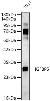
Western blot analysis of lysates from 293T cells, using IGFBP5 Rabbit pAb at 1:2000 dilution. Secondary antibody: HRP-conjugated Goat anti-Rabbit IgG (H+L) at 1:10000 dilution. Lysates/proteins: 25μg per lane. Blocking buffer: 3% nonfat dry milk in TBST. Detection: ECL Basic Kit . Exposure time: 45s.WB
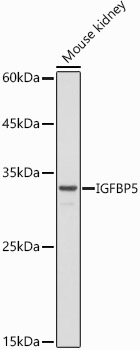
Western blot analysis of lysates from Mouse kidney, using IGFBP5 Rabbit pAb at 1:500 dilution. Secondary antibody: HRP-conjugated Goat anti-Rabbit IgG (H+L) at 1:10000 dilution. Lysates/proteins: 25μg per lane. Blocking buffer: 3% nonfat dry milk in TBST. Detection: ECL Basic Kit. Exposure time: 30s.WB
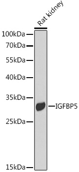
Western blot analysis of lysates from Rat kidney, using IGFBP5 Rabbit pAb at 1:1000 dilution. Secondary antibody: HRP-conjugated Goat anti-Rabbit IgG (H+L) at 1:10000 dilution. Lysates/proteins: 25μg per lane. Blocking buffer: 3% nonfat dry milk in TBST. Detection: ECL Basic Kit. Exposure time: 30s.IHC
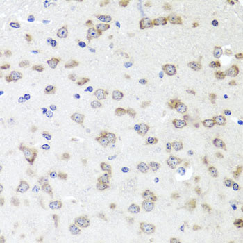
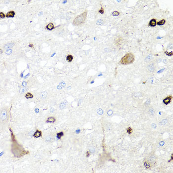
Immunohistochemistry analysis of paraffin embedded Mouse brain using IGFBP5 Rabbit pAb at dilution of 1:100 (40x lens). Microwave antigen retrieval performed with 0.01M PBS Buffer (pH 7.2) prior to IHC staining.IHC
Immunohistochemistry analysis of paraffin embedded Rat brain using IGFBP5 Rabbit pAb at dilution of 1:100 (40x lens). Microwave antigen retrieval performed with 0.01M PBS Buffer (pH 7.2) prior to IHC staining.ICC/IF
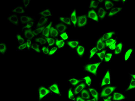
Immunofluorescence analysis of HeLa cells using IGFBP5 Rabbit pAb.Secondary antibody: Omnimabs® 488 Goat Anti-Rabbit IgG(H&L) at 1:500 dilution.| Product Name | IGFBP5 Rabbit pAb |
|---|---|
| Antibody Type | Primary Antibodies |
| Immunogen | Recombinant protein. |
| Clonality | Polyclonal |
|---|---|
| Isotype | IgG |
| Host Species | Rabbit |
| Tested Applications | ICC/IFIHCWB |
| WB:1:500-1:1000 IHC:1:50-1:200 ICC/IF:1:50-1:200 |
|
| Species Reactivity | HumanMouseRat |
| Concentration | 1mg/ml |
| Purification | Affinity purified |
| Gene Symbol | IGFBP5 |
|---|---|
| Gene Synonyms | IBP5 |
| Gene Full Name | insulin like growth factor binding protein 5 |
| Gene Summary | Enables insulin-like growth factor I binding activity and receptor ligand activity. Involved in several processes, including cellular response to cAMP; regulation of smooth muscle cell migration; and regulation of smooth muscle cell proliferation. Part of insulin-like growth factor ternary complex. Biomarker of pulmonary fibrosis. [provided by Alliance of Genome Resources, Jul 2025] |
| Alternative Names | IBP5 IGFBP5 |
| Molecular Weight(MW) | 31kDa(Observed MW 33kDa) |
| Cellular Localization | Secreted. |
WB
Western blot analysis of lysates from 293T cells, using IGFBP5 Rabbit pAb at 1:2000 dilution. Secondary antibody: HRP-conjugated Goat anti-Rabbit IgG (H+L) at 1:10000 dilution. Lysates/proteins: 25μg per lane. Blocking buffer: 3% nonfat dry milk in TBST. Detection: ECL Basic Kit . Exposure time: 45s.
WB
Western blot analysis of lysates from Mouse kidney, using IGFBP5 Rabbit pAb at 1:500 dilution. Secondary antibody: HRP-conjugated Goat anti-Rabbit IgG (H+L) at 1:10000 dilution. Lysates/proteins: 25μg per lane. Blocking buffer: 3% nonfat dry milk in TBST. Detection: ECL Basic Kit. Exposure time: 30s.
WB
Western blot analysis of lysates from Rat kidney, using IGFBP5 Rabbit pAb at 1:1000 dilution. Secondary antibody: HRP-conjugated Goat anti-Rabbit IgG (H+L) at 1:10000 dilution. Lysates/proteins: 25μg per lane. Blocking buffer: 3% nonfat dry milk in TBST. Detection: ECL Basic Kit. Exposure time: 30s.
IHC
Immunohistochemistry analysis of paraffin embedded Mouse brain using IGFBP5 Rabbit pAb at dilution of 1:100 (40x lens). Microwave antigen retrieval performed with 0.01M PBS Buffer (pH 7.2) prior to IHC staining.
IHC
Immunohistochemistry analysis of paraffin embedded Rat brain using IGFBP5 Rabbit pAb at dilution of 1:100 (40x lens). Microwave antigen retrieval performed with 0.01M PBS Buffer (pH 7.2) prior to IHC staining.
ICC/IF
Immunofluorescence analysis of HeLa cells using IGFBP5 Rabbit pAb.Secondary antibody: Omnimabs® 488 Goat Anti-Rabbit IgG(H&L) at 1:500 dilution.| Application Notes | WB:1:500-1:1000 IHC:1:50-1:200 ICC/IF:1:50-1:200 |
|---|
| Form | Liquid |
|---|---|
| Storage Instructions | Store at -20℃. Avoid freeze / thaw cycles. |
| Storage Buffer | Buffer: PBS with 0.05% proclin300, 0.05% BSA, 50% glycerol, pH7.3. |


