WB
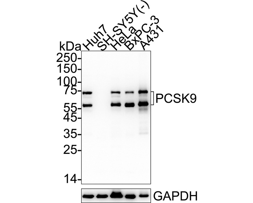
Western blot analysis of PCSK9 on different lysates with Rabbit anti-PCSK9 antibody at 1/5,000 dilution. Lane 1: Huh7 cell lysate, Lane 2: SH-SY5Y cell lysate (negative), Lane 3: HeLa cell lysate, Lane 4: BxPC-3 cell lysate, Lane 6: A431 cell lysate, Lysates/proteins at 15 µg/Lane. Exposure time: 15 seconds; 4-20% SDS-PAGE gel. Proteins were transferred to a PVDF membrane and blocked with 5% NFDM/TBST for 1 hour at room temperature. The primary antibody at 1/5,000 dilution was used in primary antibody dilution at 4℃ overnight. Goat Anti-Rabbit IgG - HRP Secondary Antibody at 1/50,000 dilution was used for 1 hour at room temperature.WB
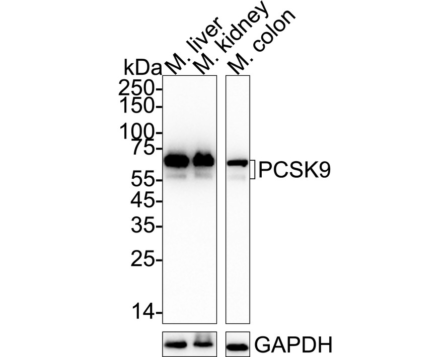
Western blot analysis of PCSK9 on different lysates with Rabbit anti-PCSK9 antibody at 1/5,000 dilution. Lane 1: Mouse liver tissue lysate, Lane 2: Mouse kidney tissue lysate, Lane 3: Mouse colon tissue lysate, Lysates/proteins at 30 µg/Lane. Exposure time: 15 seconds; 4-20% SDS-PAGE gel. Proteins were transferred to a PVDF membrane and blocked with 5% NFDM/TBST for 1 hour at room temperature. The primary antibody at 1/5,000 dilution was used in primary antibody dilution at 4℃ overnight. Goat Anti-Rabbit IgG - HRP Secondary Antibody at 1/50,000 dilution was used for 1 hour at room temperature.FC
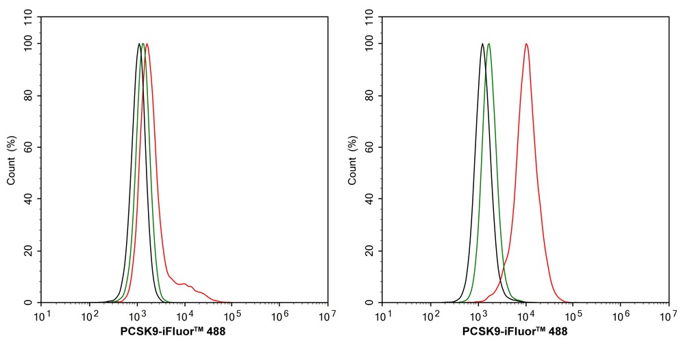
Flow cytometric analysis of SH-SY5Y (left, negative) and Huh7 (right, positive) cells labeling PCSK9. Cells were fixed and permeabilized. Then stained with the primary antibody (1/1,000) (red) compared with Rabbit IgG Isotype Control (green). After incubation of the primary antibody at +4℃ for an hour, the cells were stained with a 488 conjugate-Goat anti-Rabbit IgG Secondary antibody at 1/1,000 dilution for 30 minutes at +4℃. Unlabelled sample was used as a control (cells without incubation with primary antibody; black).IP
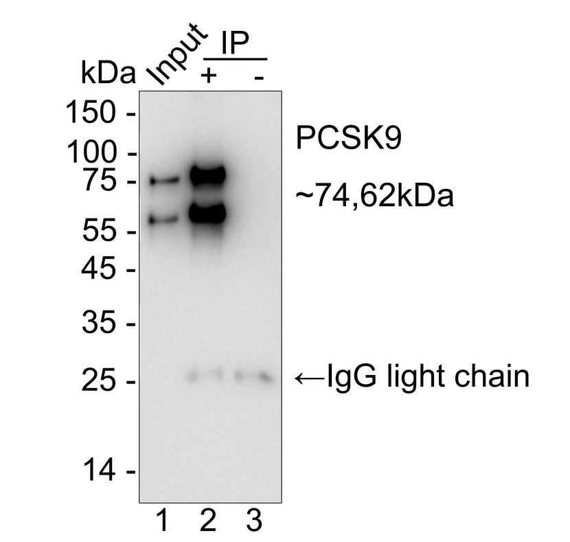
PCSK9 was immunoprecipitated from 0.2 mg Huh7 cell lysate with Rabbit anti-PCSK9 antibody at 2 µg/10 µl beads. Western blot was performed from the immunoprecipitate using Rabbit anti-PCSK9 antibody at 1/5,000 dilution. HRP Conjugated Anti-Rabbit IgG for IP Nano-secondary antibody at 1/5,000 dilution was used for 1 hour at room temperature. Lane 1: Huh7 cell lysate (input), Lane 2: Rabbit anti-PCSK9 antibody IP in Huh7 cell lysate, Lane 3: Rabbit IgG instead of Rabbit anti-PCSK9 antibody in Huh7 cell lysate, Blocking/Dilution buffer: primary antibody dilution, Exposure time: 19 seconds.| Product Name | PCSK9 Recombinant Rabbit Monoclonal Antibody |
|---|---|
| Antibody Type | Primary Antibodies |
| Immunogen | Recombinant protein within mouse PCSK9 aa 1-694. |
| Clonality | Monoclonal |
|---|---|
| Isotype | IgG |
| Host Species | Rabbit |
| Tested Applications | FCIPWB |
| WB:1:5000 FC:1:1000 IP:1-2μg/sample |
|
| Species Reactivity | HumanMouse |
| Concentration | 1mg/ml |
| Purification | Protein A |
| Gene Symbol | PCSK9 |
|---|---|
| Gene Synonyms | FH3 PC9 FHCL3 NARC1 LDLCQ1 NARC-1 HCHOLA3 |
| Gene Full Name | proprotein convertase subtilisin/kexin type 9 |
| Gene Summary | This gene encodes a member of the subtilisin-like proprotein convertase family, which includes proteases that process protein and peptide precursors trafficking through regulated or constitutive branches of the secretory pathway. The encoded protein undergoes an autocatalytic processing event with its prosegment in the ER and is constitutively secreted as an inactive protease into the extracellular matrix and trans-Golgi network. It is expressed in liver, intestine and kidney tissues and escorts specific receptors for lysosomal degradation. It plays a role in cholesterol and fatty acid metabolism. Mutations in this gene have been associated with autosomal dominant familial hypercholesterolemia. Alternative splicing results in multiple transcript variants. [provided by RefSeq, Feb 2014] |
| Alternative Names | Convertase subtilisin/kexin type 9 preproprotein antibody FH3 antibody HCHOLA3 antibody Hypercholesterolemia autosomal dominant 3 antibody LDLCQ1 antibody NARC 1 antibody NARC-1 antibody NARC1 antibody Neural apoptosis regulated convertase 1 antibody Neural apoptosis-regulated convertase 1 antibody Convertase subtilisin/kexin type 9 preproprotein antibody FH3 antibody HCHOLA3 antibody Hypercholesterolemia autosomal dominant 3 antibody LDLCQ1 antibody NARC 1 antibody NARC-1 antibody NARC1 antibody Neural apoptosis regulated convertase 1 antibody Neural apoptosis-regulated convertase 1 antibody PC 9 antibody PC9 antibody PCSK 9 antibody PCSK9 antibody PCSK9_HUMAN antibody Proprotein convertase 9 antibody Proprotein convertase PC9 antibody Proprotein convertase subtilisin/kexin type 9 antibody PSEC0052 antibody Subtilisin/kexin like protease PC9 antibody Subtilisin/kexin-like protease PC9 antibody |
| Molecular Weight(MW) | 74 kDa(Observed band size: 62/74 kDa) |
| Cellular Localization | Cytoplasm, Secreted, Endosome, Lysosome, Cell surface, Endoplasmic reticulum, Golgi apparatus. |
WB
Western blot analysis of PCSK9 on different lysates with Rabbit anti-PCSK9 antibody at 1/5,000 dilution. Lane 1: Huh7 cell lysate, Lane 2: SH-SY5Y cell lysate (negative), Lane 3: HeLa cell lysate, Lane 4: BxPC-3 cell lysate, Lane 6: A431 cell lysate, Lysates/proteins at 15 µg/Lane. Exposure time: 15 seconds; 4-20% SDS-PAGE gel. Proteins were transferred to a PVDF membrane and blocked with 5% NFDM/TBST for 1 hour at room temperature. The primary antibody at 1/5,000 dilution was used in primary antibody dilution at 4℃ overnight. Goat Anti-Rabbit IgG - HRP Secondary Antibody at 1/50,000 dilution was used for 1 hour at room temperature.
WB
Western blot analysis of PCSK9 on different lysates with Rabbit anti-PCSK9 antibody at 1/5,000 dilution. Lane 1: Mouse liver tissue lysate, Lane 2: Mouse kidney tissue lysate, Lane 3: Mouse colon tissue lysate, Lysates/proteins at 30 µg/Lane. Exposure time: 15 seconds; 4-20% SDS-PAGE gel. Proteins were transferred to a PVDF membrane and blocked with 5% NFDM/TBST for 1 hour at room temperature. The primary antibody at 1/5,000 dilution was used in primary antibody dilution at 4℃ overnight. Goat Anti-Rabbit IgG - HRP Secondary Antibody at 1/50,000 dilution was used for 1 hour at room temperature.
FC
Flow cytometric analysis of SH-SY5Y (left, negative) and Huh7 (right, positive) cells labeling PCSK9. Cells were fixed and permeabilized. Then stained with the primary antibody (1/1,000) (red) compared with Rabbit IgG Isotype Control (green). After incubation of the primary antibody at +4℃ for an hour, the cells were stained with a 488 conjugate-Goat anti-Rabbit IgG Secondary antibody at 1/1,000 dilution for 30 minutes at +4℃. Unlabelled sample was used as a control (cells without incubation with primary antibody; black).
IP
PCSK9 was immunoprecipitated from 0.2 mg Huh7 cell lysate with Rabbit anti-PCSK9 antibody at 2 µg/10 µl beads. Western blot was performed from the immunoprecipitate using Rabbit anti-PCSK9 antibody at 1/5,000 dilution. HRP Conjugated Anti-Rabbit IgG for IP Nano-secondary antibody at 1/5,000 dilution was used for 1 hour at room temperature. Lane 1: Huh7 cell lysate (input), Lane 2: Rabbit anti-PCSK9 antibody IP in Huh7 cell lysate, Lane 3: Rabbit IgG instead of Rabbit anti-PCSK9 antibody in Huh7 cell lysate, Blocking/Dilution buffer: primary antibody dilution, Exposure time: 19 seconds.| Application Notes | WB:1:5000 FC:1:1000 IP:1-2μg/sample |
|---|
| Form | Liquid |
|---|---|
| Storage Instructions | Store at +4℃ after thawing. Aliquot store at -20℃. Avoid repeated freeze / thaw cycles. |
| Storage Buffer | PBS (pH7.4), 0.1% BSA, 40% Glycerol. Preservative: 0.05% Sodium Azide. |


