WB
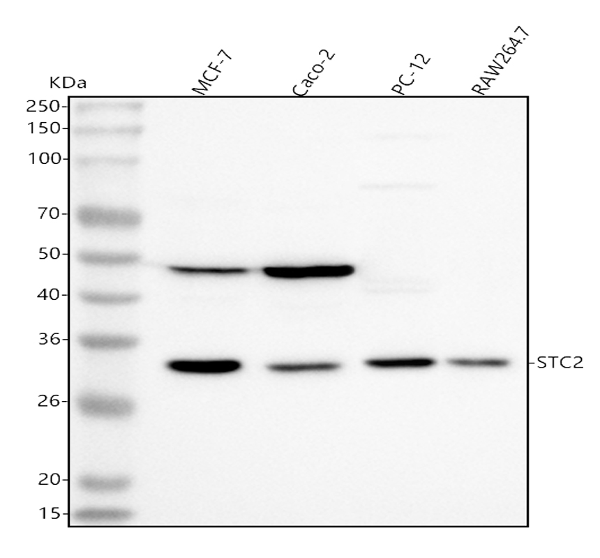
Western blot analysis of Stanniocalcin 2/STC2 using anti-Stanniocalcin 2/STC2 antibody. The sample well of each lane was loaded with 30 ug of sample under reducing conditions. Lane 1: human MCF-7 whole cell lysates, Lane 2: human Caco-2 whole cell lysates, Lane 3: rat PC-12 whole cell lysates, Lane 4: mouse RAW264.7 whole cell lysates. After electrophoresis, proteins were transferred to a membrane. Then the membrane was incubated with rabbit anti-Stanniocalcin 2/STC2 antigen affinity purified polyclonal antibody at a dilution of 1:1000 and probed with a goat anti-rabbit IgG-HRP secondary antibody. The signal is developed using ECL Plus Western Blotting Substrate.IHC
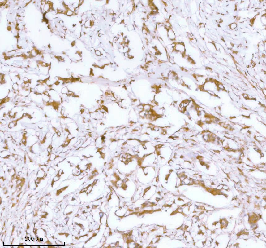
IHC analysis of Stanniocalcin 2/STC2 using anti-Stanniocalcin 2/STC2 antibody. Stanniocalcin 2/STC2 was detected in a paraffin-embedded section of human breast cancer tissue. The tissue section was incubated with rabbit anti-Stanniocalcin 2/STC2 Antibody at a dilution of 1:200 and developed using HRP Conjugated Rabbit IgG Super Vision Assay Kit with DAB as the chromogen.IHC
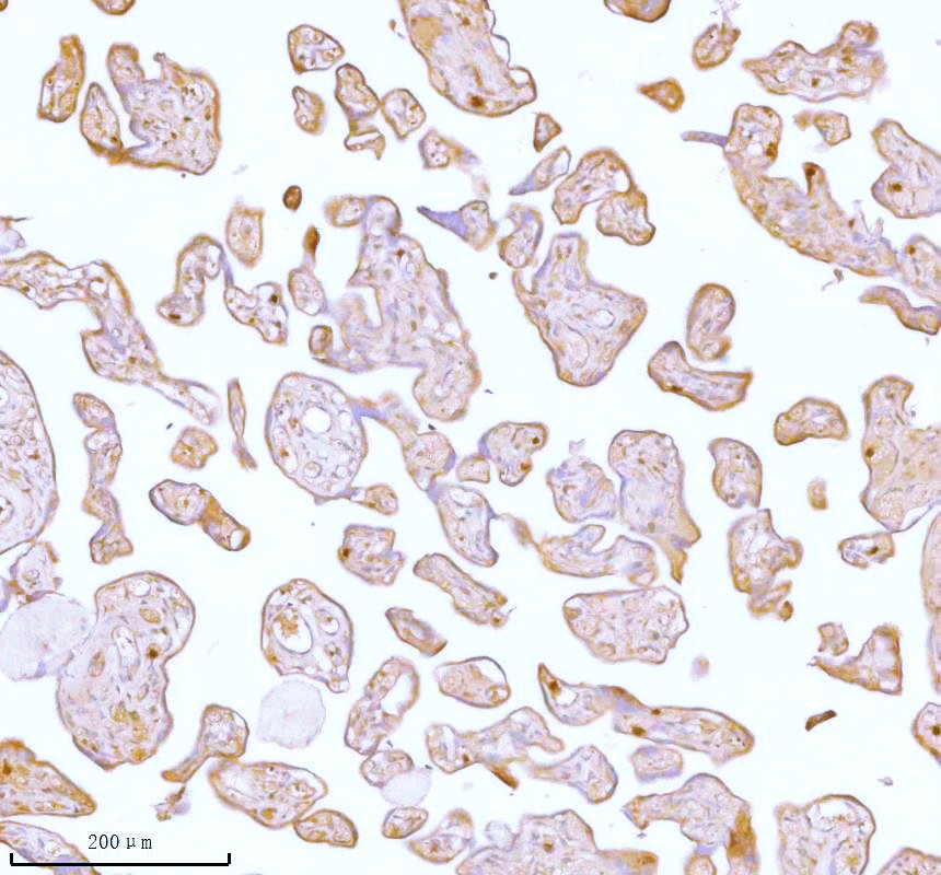
IHC analysis of Stanniocalcin 2/STC2 using anti-Stanniocalcin 2/STC2 antibody. Stanniocalcin 2/STC2 was detected in a paraffin-embedded section of human placenta tissue. The tissue section was incubated with rabbit anti-Stanniocalcin 2/STC2 Antibody at a dilution of 1:200 and developed using HRP Conjugated Rabbit IgG Super Vision Assay Kit with DAB as the chromogen.ICC/IF
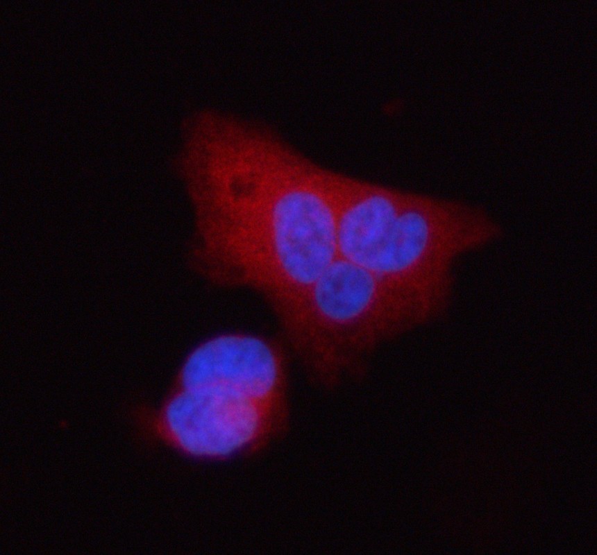
ICC/IF analysis of Stanniocalcin 2/STC2 using anti-Stanniocalcin 2/STC2 antibody. Stanniocalcin 2/STC2 was detected in an immunocytochemical section of PC-3 cells. The section was incubated with rabbit anti-Stanniocalcin 2/STC2 Antibody at a dilution of 1:100. Cy3-conjugated Anti-rabbit IgG Secondary Antibody (red) was used as secondary antibody. The section was counterstained with DAPI (Blue).IF-P
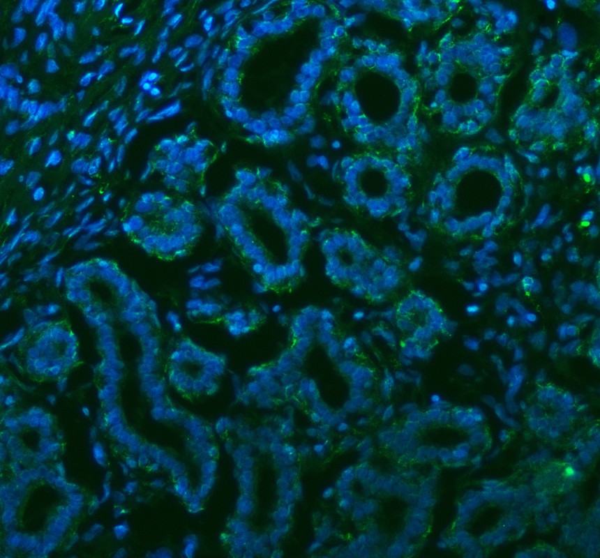
IF analysis of Stanniocalcin 2/STC2 using anti-Stanniocalcin 2/STC2 antibody. Stanniocalcin 2/STC2 was detected in a paraffin-embedded section of human lung cancer tissue. The tissue section was incubated with rabbit anti-Stanniocalcin 2/STC2 Antibody at a dilution of 1:100. Fluoro488-conjugated Anti-rabbit IgG Secondary Antibody (green) was used as secondary antibody. The section was counterstained with DAPI (Blue).IF-P
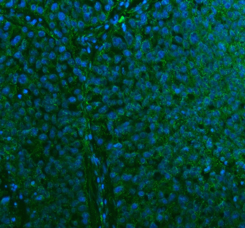
IF analysis of Stanniocalcin 2/STC2 using anti-Stanniocalcin 2/STC2 antibody. Stanniocalcin 2/STC2 was detected in a paraffin-embedded section of human breast cancer tissue. The tissue section was incubated with rabbit anti-Stanniocalcin 2/STC2 Antibody at a dilution of 1:100. Fluoro488-conjugated Anti-rabbit IgG Secondary Antibody (green) was used as secondary antibody. The section was counterstained with DAPI (Blue).FC
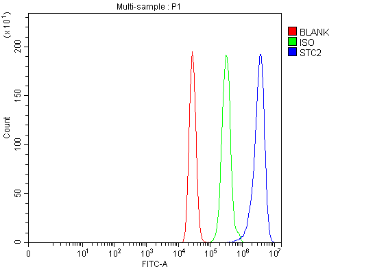
Flow Cytometry analysis of MCF-7 cells using anti-Stanniocalcin 2/STC2 antibody. Overlay histogram showing MCF-7 cells stained with A05017 (Blue line). The cells were fixed with 4% paraformaldehyde and blocked with 10% normal goat serum. And then incubated with rabbit anti-Stanniocalcin 2/STC2 Antibody at 1:100 dilution for 30 min at 20°C. Fluoro488 conjugated goat anti-rabbit IgG was used as secondary antibody at 1:100 dilution for 30 minutes at 20°C. Isotype control antibody (Green line) was rabbit IgG at 1:100 dilution used under the same conditions. Unlabelled sample (Red line) was also used as a control.| Product Name | Rabbit polyclonal antibody to Stanniocalcin 2/STC2 |
|---|---|
| Antibody Type | Primary Antibodies |
| Immunogen | E.coli-derived human Stanniocalcin 2/STC2 recombinant protein (Position: A51-Q226). Human STC2 shares 90.3% and 89.8% amino acid (aa) sequence identity with mouse and rat STC2, respectively. |
| Clonality | Polyclonal |
|---|---|
| Isotype | IgG |
| Host Species | Rabbit |
| Tested Applications | FCICC/IFIF-PIHCWB |
| WB:1:500-1:2000 IHC:1:50-1:400 ICC/IF:1:50-1:400 IF-P:1:50-1:400 FC:1:50-1:200 |
|
| Species Reactivity | HumanMouseRat |
| Concentration | 0.5mg/ml |
| Purification | Affinity purified |
| Gene Symbol | STC2 |
|---|---|
| Gene Synonyms | STC-2 STCRP |
| Gene Full Name | stanniocalcin 2 |
| Gene Summary | This gene encodes a secreted, homodimeric glycoprotein that is expressed in a wide variety of tissues and may have autocrine or paracrine functions. The encoded protein has 10 of its 15 cysteine residues conserved among stanniocalcin family members and is phosphorylated by casein kinase 2 exclusively on its serine residues. Its C-terminus contains a cluster of histidine residues which may interact with metal ions. The protein may play a role in the regulation of renal and intestinal calcium and phosphate transport, cell metabolism, or cellular calcium/phosphate homeostasis. Constitutive overexpression of human stanniocalcin 2 in mice resulted in pre- and postnatal growth restriction, reduced bone and skeletal muscle growth, and organomegaly. Expression of this gene is induced by estrogen and altered in some breast cancers. [provided by RefSeq, Jul 2008] |
| Alternative Names | stanniocalcin 2 Stanniocalcin related protein STC 2 STC related protein STC2 STCRP |
| Molecular Weight(MW) | 33 kDa |
WB
Western blot analysis of Stanniocalcin 2/STC2 using anti-Stanniocalcin 2/STC2 antibody. The sample well of each lane was loaded with 30 ug of sample under reducing conditions. Lane 1: human MCF-7 whole cell lysates, Lane 2: human Caco-2 whole cell lysates, Lane 3: rat PC-12 whole cell lysates, Lane 4: mouse RAW264.7 whole cell lysates. After electrophoresis, proteins were transferred to a membrane. Then the membrane was incubated with rabbit anti-Stanniocalcin 2/STC2 antigen affinity purified polyclonal antibody at a dilution of 1:1000 and probed with a goat anti-rabbit IgG-HRP secondary antibody. The signal is developed using ECL Plus Western Blotting Substrate.
IHC
IHC analysis of Stanniocalcin 2/STC2 using anti-Stanniocalcin 2/STC2 antibody. Stanniocalcin 2/STC2 was detected in a paraffin-embedded section of human breast cancer tissue. The tissue section was incubated with rabbit anti-Stanniocalcin 2/STC2 Antibody at a dilution of 1:200 and developed using HRP Conjugated Rabbit IgG Super Vision Assay Kit with DAB as the chromogen.
IHC
IHC analysis of Stanniocalcin 2/STC2 using anti-Stanniocalcin 2/STC2 antibody. Stanniocalcin 2/STC2 was detected in a paraffin-embedded section of human placenta tissue. The tissue section was incubated with rabbit anti-Stanniocalcin 2/STC2 Antibody at a dilution of 1:200 and developed using HRP Conjugated Rabbit IgG Super Vision Assay Kit with DAB as the chromogen.
ICC/IF
ICC/IF analysis of Stanniocalcin 2/STC2 using anti-Stanniocalcin 2/STC2 antibody. Stanniocalcin 2/STC2 was detected in an immunocytochemical section of PC-3 cells. The section was incubated with rabbit anti-Stanniocalcin 2/STC2 Antibody at a dilution of 1:100. Cy3-conjugated Anti-rabbit IgG Secondary Antibody (red) was used as secondary antibody. The section was counterstained with DAPI (Blue).
IF-P
IF analysis of Stanniocalcin 2/STC2 using anti-Stanniocalcin 2/STC2 antibody. Stanniocalcin 2/STC2 was detected in a paraffin-embedded section of human lung cancer tissue. The tissue section was incubated with rabbit anti-Stanniocalcin 2/STC2 Antibody at a dilution of 1:100. Fluoro488-conjugated Anti-rabbit IgG Secondary Antibody (green) was used as secondary antibody. The section was counterstained with DAPI (Blue).
IF-P
IF analysis of Stanniocalcin 2/STC2 using anti-Stanniocalcin 2/STC2 antibody. Stanniocalcin 2/STC2 was detected in a paraffin-embedded section of human breast cancer tissue. The tissue section was incubated with rabbit anti-Stanniocalcin 2/STC2 Antibody at a dilution of 1:100. Fluoro488-conjugated Anti-rabbit IgG Secondary Antibody (green) was used as secondary antibody. The section was counterstained with DAPI (Blue).
FC
Flow Cytometry analysis of MCF-7 cells using anti-Stanniocalcin 2/STC2 antibody. Overlay histogram showing MCF-7 cells stained with A05017 (Blue line). The cells were fixed with 4% paraformaldehyde and blocked with 10% normal goat serum. And then incubated with rabbit anti-Stanniocalcin 2/STC2 Antibody at 1:100 dilution for 30 min at 20°C. Fluoro488 conjugated goat anti-rabbit IgG was used as secondary antibody at 1:100 dilution for 30 minutes at 20°C. Isotype control antibody (Green line) was rabbit IgG at 1:100 dilution used under the same conditions. Unlabelled sample (Red line) was also used as a control.| Application Notes | WB:1:500-1:2000 IHC:1:50-1:400 ICC/IF:1:50-1:400 IF-P:1:50-1:400 FC:1:50-1:200 |
|---|
| Form | Liquid |
|---|---|
| Storage Instructions | 12 months from date of receipt, -20℃ as supplied. 6 months 2 to 8℃ after reconstitution. Avoid repeated freezing and thawing. |
| Storage Buffer | 500ug/ml antibody with PBS, 0.02% NaN3, 1 mg/ml BSA and 50% glycerol. |


