WB
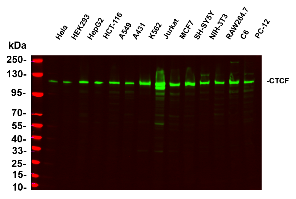
Various whole cell lysates were separated by 4-20% SDS-PAGE, and the primary antibody was used at 4~C, over night with a 1:5000 dilution . The Dylight 800-conjugated Goat anti-Rabbit antibody was used to detect the antibody. Lane1: Hela - Human cervical cancer, Lane2: HEK293 - Human normal embryonic kidney, Lane3: HepG2 - Human hepatocellular carcinoma, Lane4: HCT-116 - Human colon cancer, Lane5: A549 - Human lung adenocarcinoma, Lane6: A431 - Human skin squamous cell carcinoma, Lane7: K562 - Human chronic myeloid leukemia, Lane8: Jurkat - Human acute T cell leukemia cells, Lane9: MCF7 - Human breast cancer, Lane10: SH-SY5Y - Human neuroblastoma cells, Lane11: NIH-3T3 - NIH mouse fibroblasts, Lane12: RAW264.7 - Mouse mononuclear macrophage leukemia cells, Lane13: C6 - Rat glioma cells, Lane14: PC-12 - Pheochromocytoma in rats.IHC
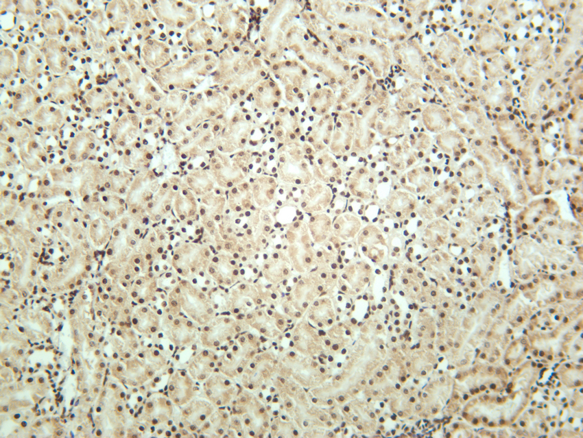
Mouse kidney was stained with anti-CTCF rabbit antibody.IHC
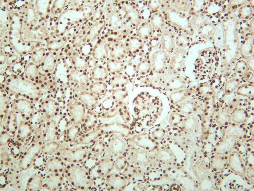
Rat kidney was stained with anti-CTCF rabbit antibody.ICC.IF
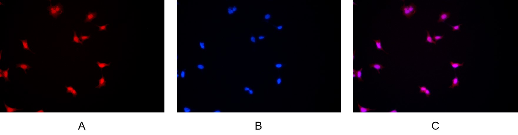
Immunofluorescence analysis of HEK293. Picture A: CTCF antibody (red). Picture B: DAPI (blue). Picture C: Merge of A+B.| Product Name | CTCF Rabbit mAb |
|---|---|
| Antibody Type | Primary Antibodies |
| Clonality | Monoclonal |
|---|---|
| Isotype | IgG |
| Host Species | Rabbit |
| Tested Applications | ELISAICC/IFIHCWB |
| WB:1:2000-1:10000 IHC:1:2000-1:10000 ICC/IF:1:200-1:1000 ELISA:1:1000-1:10000 |
|
| Species Reactivity | HumanMouseRat |
| Concentration | 1mg/ml |
| Purification | Protein A |
| Gene Symbol | CTCF |
|---|---|
| Gene Synonyms | MRD21 FAP108 CFAP108 |
| Gene Full Name | CCCTC-binding factor |
| Gene Summary | This gene is a member of the BORIS + CTCF gene family and encodes a transcriptional regulator protein with 11 highly conserved zinc finger (ZF) domains. This nuclear protein is able to use different combinations of the ZF domains to bind different DNA target sequences and proteins. Depending upon the context of the site, the protein can bind a histone acetyltransferase (HAT)-containing complex and function as a transcriptional activator or bind a histone deacetylase (HDAC)-containing complex and function as a transcriptional repressor. If the protein is bound to a transcriptional insulator element, it can block communication between enhancers and upstream promoters, thereby regulating imprinted expression. Mutations in this gene have been associated with invasive breast cancers, prostate cancers, and Wilms' tumors. Alternatively spliced transcript variants encoding different isoforms have been found for this gene. [provided by RefSeq, Jul 2010] |
| Alternative Names | CTCF Transcriptional repressor CTCF 11-zinc finger protein CCCTC-binding factor CTCFL paralog |
| Molecular Weight(MW) | 83kDa(140kDa) |
| Cellular Localization | Nucleus. |
WB
Various whole cell lysates were separated by 4-20% SDS-PAGE, and the primary antibody was used at 4~C, over night with a 1:5000 dilution . The Dylight 800-conjugated Goat anti-Rabbit antibody was used to detect the antibody. Lane1: Hela - Human cervical cancer, Lane2: HEK293 - Human normal embryonic kidney, Lane3: HepG2 - Human hepatocellular carcinoma, Lane4: HCT-116 - Human colon cancer, Lane5: A549 - Human lung adenocarcinoma, Lane6: A431 - Human skin squamous cell carcinoma, Lane7: K562 - Human chronic myeloid leukemia, Lane8: Jurkat - Human acute T cell leukemia cells, Lane9: MCF7 - Human breast cancer, Lane10: SH-SY5Y - Human neuroblastoma cells, Lane11: NIH-3T3 - NIH mouse fibroblasts, Lane12: RAW264.7 - Mouse mononuclear macrophage leukemia cells, Lane13: C6 - Rat glioma cells, Lane14: PC-12 - Pheochromocytoma in rats.
IHC
Mouse kidney was stained with anti-CTCF rabbit antibody.
IHC
Rat kidney was stained with anti-CTCF rabbit antibody.
ICC.IF
Immunofluorescence analysis of HEK293. Picture A: CTCF antibody (red). Picture B: DAPI (blue). Picture C: Merge of A+B.| Application Notes | WB:1:2000-1:10000 IHC:1:2000-1:10000 ICC/IF:1:200-1:1000 ELISA:1:1000-1:10000 |
|---|
| Form | Liquid |
|---|---|
| Storage Instructions | -15°C to -25°C/1 year(Do not lower than -25°C) |
| Storage Buffer | PBS, 50% glycerol, 0.05% Proclin 300, 0.05%BSA |


