WB
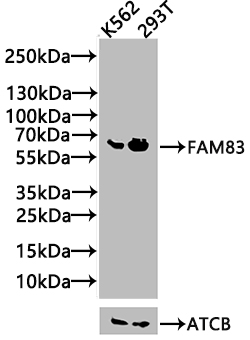
Positive WB detected in: K562 whole cell lysate(20µg), 293T whole cell lysate(20µg), All lanes: FAM83D antibody at 1:1000, Secondary Goat polyclonal to rabbit IgG at 1/20000 dilution.IHC
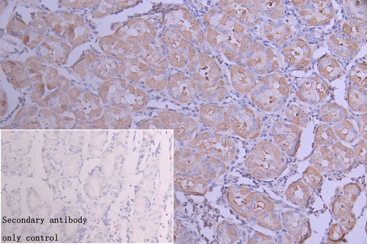
IHC image of FAM83D Antibody diluted at 1:300 and staining in paraffin-embedded human stomach tissue performed on a Leica BondTM system. After dewaxing and hydration, antigen retrieval was mediated by high pressure in a citrate buffer (pH 6.0). Section was blocked with 10% normal goat serum 30min at RT. Then primary antibody (1% BSA) was incubated at 4°C overnight. The primary is detected by a Goat anti-rabbit polymer IgG labeled by HRP and visualized using 0.05% DAB. Secondary antibody only control: uses 1% BSA instead of primary antibody.IHC
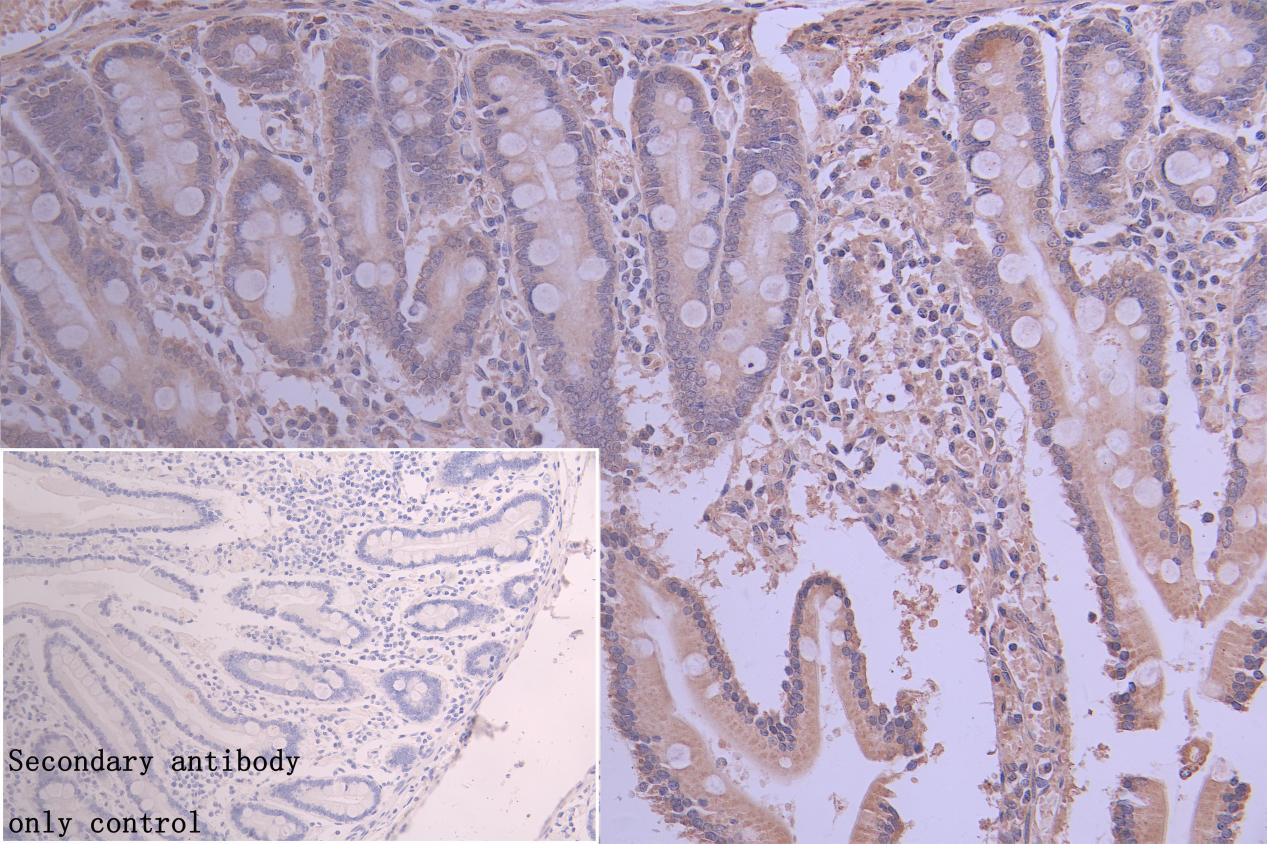
IHC image ofFAM83D Antibody diluted at 1:300 and staining in paraffin-embedded human gastric tissue performed on a Leica BondTM system. After dewaxing and hydration, antigen retrieval was mediated by high pressure in a citrate buffer (pH 6.0). Section was blocked with 10% normal goat serum 30min at RT. Then primary antibody (1% BSA) was incubated at 4°C overnight. The primary is detected by a Goat anti-rabbit polymer IgG labeled by HRP and visualized using 0.05% DAB. Secondary antibody only control: uses 1% BSA instead of primary antibody.| Product Name | Rabbit anti-FAM83D Polyclonal antibody |
|---|---|
| Antibody Type | Primary Antibodies |
| Immunogen | Recombinant Human Protein FAM83D protein (339-470AA) |
| Clonality | Polyclonal |
|---|---|
| Isotype | IgG |
| Host Species | Rabbit |
| Tested Applications | IHCWB |
| WB:1:500-1:5000 IHC:1:100-1:300 |
|
| Species Reactivity | Human |
| Concentration | 1mg/ml |
| Purification | Affinity purified |
| Gene Symbol | SACK1D |
|---|---|
| Gene Synonyms | CHICA FAM83D C20orf129 dJ616B8.3 |
| Gene Full Name | scaffolding CK1 anchoring protein D |
| Gene Summary | Enables kinesin binding activity; microtubule binding activity; and protein kinase binding activity. Involved in several processes, including positive regulation of cell cycle G1/S phase transition; protein localization to mitotic spindle; and regulation of intracellular signal transduction. Located in cytosol; intercellular bridge; and mitotic spindle pole. [provided by Alliance of Genome Resources, Jul 2025] |
| Alternative Names | 2310007D09Rik antibody BB104611 antibody C20orf129 antibody CHICA antibody dJ616B8.3 antibody FA83D antibody FA83D_HUMAN antibody FAM83D antibody Family with sequence similarity 83 member D antibody FLJ38341 antibody MGC92947 antibody Protein FAM83D antibody RGD1565583 antibody RP23-119H5.1 antibody Spindle protein CHICA antibody |
| Molecular Weight(MW) | 65 kDa |
| Cellular Localization | Cytoplasm. Cytoplasm, cytoskeleton, spindle. Cytoplasm, cytoskeleton, spindle pole. |
WB
Positive WB detected in: K562 whole cell lysate(20µg), 293T whole cell lysate(20µg), All lanes: FAM83D antibody at 1:1000, Secondary Goat polyclonal to rabbit IgG at 1/20000 dilution.
IHC
IHC image of FAM83D Antibody diluted at 1:300 and staining in paraffin-embedded human stomach tissue performed on a Leica BondTM system. After dewaxing and hydration, antigen retrieval was mediated by high pressure in a citrate buffer (pH 6.0). Section was blocked with 10% normal goat serum 30min at RT. Then primary antibody (1% BSA) was incubated at 4°C overnight. The primary is detected by a Goat anti-rabbit polymer IgG labeled by HRP and visualized using 0.05% DAB. Secondary antibody only control: uses 1% BSA instead of primary antibody.
IHC
IHC image ofFAM83D Antibody diluted at 1:300 and staining in paraffin-embedded human gastric tissue performed on a Leica BondTM system. After dewaxing and hydration, antigen retrieval was mediated by high pressure in a citrate buffer (pH 6.0). Section was blocked with 10% normal goat serum 30min at RT. Then primary antibody (1% BSA) was incubated at 4°C overnight. The primary is detected by a Goat anti-rabbit polymer IgG labeled by HRP and visualized using 0.05% DAB. Secondary antibody only control: uses 1% BSA instead of primary antibody.| Application Notes | WB:1:500-1:5000 IHC:1:100-1:300 |
|---|
| Form | Liquid |
|---|---|
| Storage Instructions | Upon receipt, store at -20°C or -80°C. Avoid repeated freeze. |
| Storage Buffer | 0.03% Proclin 300 Constituents: 50% Glycerol, 0.01M PBS, PH 7.4 |


