WB
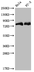
Positive WB detected in: Hela whole cell lysate, PC3 whole cell lysate, All lanes: SENP1 antibody at 1:2000, Secondary Goat polyclonal to rabbit IgG at 1/50000 dilution.IHC
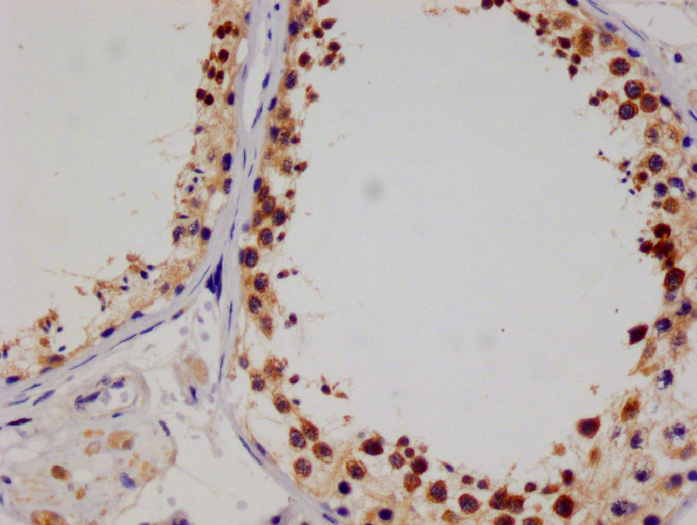
IHC image of SENP1 Antibody diluted at 1:100 and staining in paraffin-embedded human testis tissue performed on a Leica BondTM system. After dewaxing and hydration, antigen retrieval was mediated by high pressure in a citrate buffer (pH 6.0). Section was blocked with 10% normal goat serum 30min at RT. Then primary antibody (1% BSA) was incubated at 4°C overnight. The primary is detected by a Goat anti-rabbit polymer IgG labeled by HRP and visualized using 0.05% DAB.ICC/IF
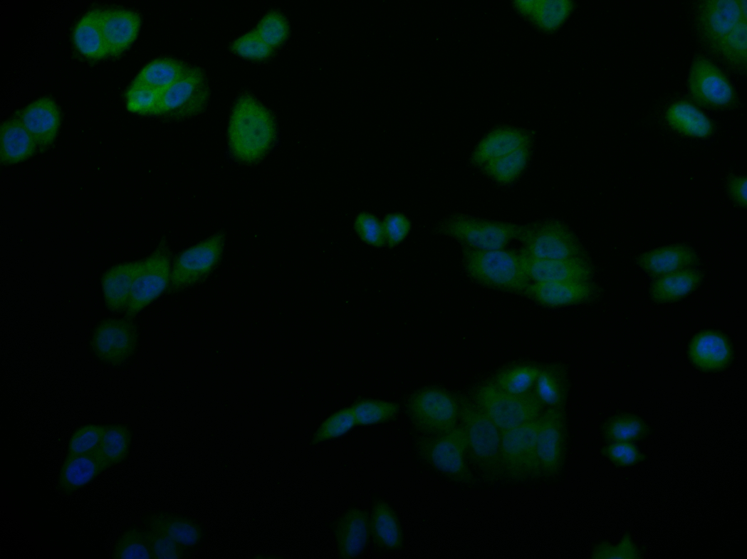
Immunofluorescence staining of PC-3 cell with SENP1 Antibody at 1:50, counter-stained with DAPI. The cells were fixed in 4% formaldehyde and blocked in 10% normal Goat Serum. The cells were then incubated with the antibody overnight at 4°C. The secondary antibody was Alexa Fluor 527-congugated AffiniPure Goat Anti-Rabbit IgG(H+L).FC
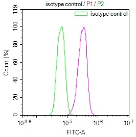
Overlay Peak curve showing K562 cells stained with SENP1 Antibody (red line) at 1:100. The cells were fixed in 4% formaldehyde and permeated by 0.2% TritonX-100. Then 10% normal goat serum to block non-specific protein-protein interactions followed by the antibody (1ug/1*106cells) for 45min at 4℃. The secondary antibody used was FITC-conjugated Goat Anti-rabbit IgG(H+L) at 1:200 dilution for 35min at 4℃.Control antibody (green line) was rabbit IgG (1ug/1*106cells) used under the same conditions. Acquisition of >10,000 events was performed.| Product Name | SENP1 Recombinant Monoclonal Antibody |
|---|---|
| Antibody Type | Primary Antibodies |
| Immunogen | A synthesized peptide derived from human SENP1. |
| Clonality | Monoclonal |
|---|---|
| Isotype | IgG |
| Host Species | Rabbit |
| Tested Applications | FCICC/IFIHCWB |
| WB:1:500-1:2000 IHC:1:50-1:200 ICC/IF:1:50-1:200 FC:1:50-1:200 |
|
| Species Reactivity | Human |
| Concentration | 1mg/ml |
| Purification | Affinity purified |
| Gene Symbol | SENP1 |
|---|---|
| Gene Synonyms | SuPr-2 |
| Gene Full Name | SUMO specific peptidase 1 |
| Gene Summary | This gene encodes a cysteine protease that specifically targets members of the small ubiquitin-like modifier (SUMO) protein family. This protease regulates SUMO pathways by deconjugating sumoylated proteins. This protease also functions to process the precursor SUMO proteins into their mature form. Alternate splicing results in multiple transcript variants. [provided by RefSeq, Jun 2012] |
| Alternative Names | Sentrin-specific protease 1 (EC 3.4.22.-) (Sentrin/SUMO-specific protease SENP1) SENP1 |
| Molecular Weight(MW) | 74 kDa |
| Cellular Localization | Nucleus. Cytoplasm. Note=Shuttles between cytoplasm and nucleus. |
WB
Positive WB detected in: Hela whole cell lysate, PC3 whole cell lysate, All lanes: SENP1 antibody at 1:2000, Secondary Goat polyclonal to rabbit IgG at 1/50000 dilution.
IHC
IHC image of SENP1 Antibody diluted at 1:100 and staining in paraffin-embedded human testis tissue performed on a Leica BondTM system. After dewaxing and hydration, antigen retrieval was mediated by high pressure in a citrate buffer (pH 6.0). Section was blocked with 10% normal goat serum 30min at RT. Then primary antibody (1% BSA) was incubated at 4°C overnight. The primary is detected by a Goat anti-rabbit polymer IgG labeled by HRP and visualized using 0.05% DAB.
ICC/IF
Immunofluorescence staining of PC-3 cell with SENP1 Antibody at 1:50, counter-stained with DAPI. The cells were fixed in 4% formaldehyde and blocked in 10% normal Goat Serum. The cells were then incubated with the antibody overnight at 4°C. The secondary antibody was Alexa Fluor 527-congugated AffiniPure Goat Anti-Rabbit IgG(H+L).
FC
Overlay Peak curve showing K562 cells stained with SENP1 Antibody (red line) at 1:100. The cells were fixed in 4% formaldehyde and permeated by 0.2% TritonX-100. Then 10% normal goat serum to block non-specific protein-protein interactions followed by the antibody (1ug/1*106cells) for 45min at 4℃. The secondary antibody used was FITC-conjugated Goat Anti-rabbit IgG(H+L) at 1:200 dilution for 35min at 4℃.Control antibody (green line) was rabbit IgG (1ug/1*106cells) used under the same conditions. Acquisition of >10,000 events was performed.| Application Notes | WB:1:500-1:2000 IHC:1:50-1:200 ICC/IF:1:50-1:200 FC:1:50-1:200 |
|---|
| Form | Liquid |
|---|---|
| Storage Instructions | Upon receipt, store at -20°C or -80°C. Avoid repeated freeze. |
| Storage Buffer | 0.03% Proclin 300 Constituents: 50% Glycerol, 0.01M PBS, PH 7.4 |


