WB
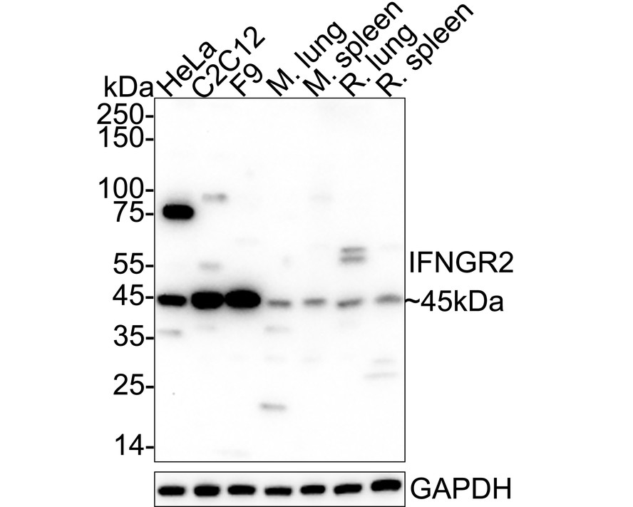
Western blot analysis of IFNGR2 on different lysates with Rabbit anti-IFNGR2 antibody at 1/1,000 dilution. Lane 1: HeLa cell lysate (20 µg/Lane), Lane 2: C2C12 cell lysate (20 µg/Lane), Lane 3: F9 cell lysate (20 µg/Lane), Lane 4: Mouse lung tissue lysate (40 µg/Lane), Lane 5: Mouse spleen tissue lysate (40 µg/Lane), Lane 6: Rat lung tissue lysate (40 µg/Lane), Lane 7: Rat spleen tissue lysate (40 µg/Lane), Exposure time: 1 minute; 4-20% SDS-PAGE gel. Proteins were transferred to a PVDF membrane and blocked with 5% NFDM/TBST for 1 hour at room temperature. The primary antibody at 1/1,000 dilution was used in 5% NFDM/TBST at 4℃ overnight. Goat Anti-Rabbit IgG - HRP Secondary Antibody at 1/50,000 dilution was used for 1 hour at room temperature.IHC
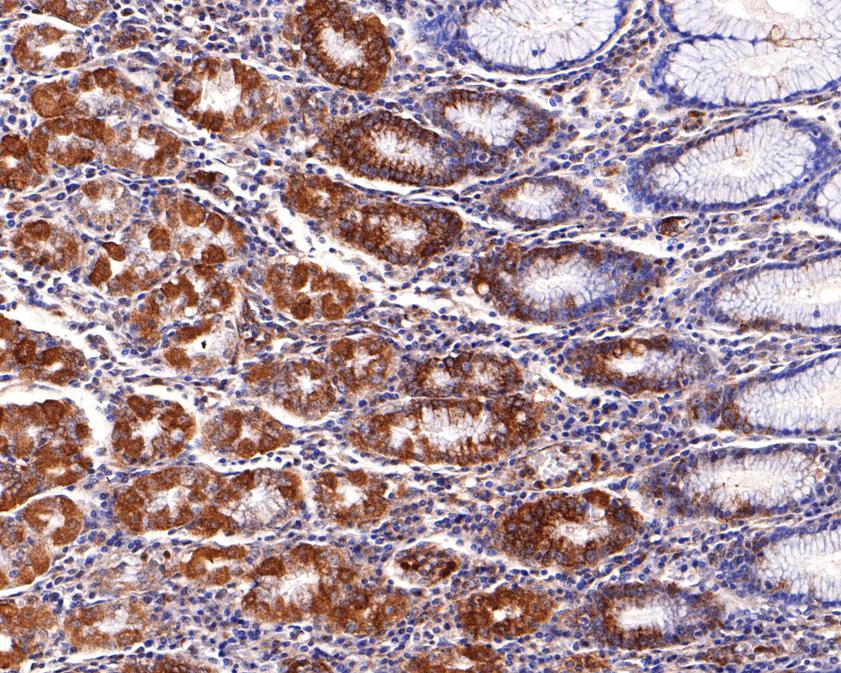
Immunohistochemical analysis of paraffin-embedded human stomach tissue using anti-IFNGR2 antibody. The section was pre-treated using heat mediated antigen retrieval with Tris-EDTA buffer (pH 9.0) for 20 minutes.The tissues were blocked in 1% BSA for 30 minutes at room temperature, washed with ddH2O and PBS, and then probed with the primary antibody (1/600) for 30 minutes at room temperature. The detection was performed using an HRP conjugated compact polymer system. DAB was used as the chromogen. Tissues were counterstained with hematoxylin and mounted with DPX.IHC
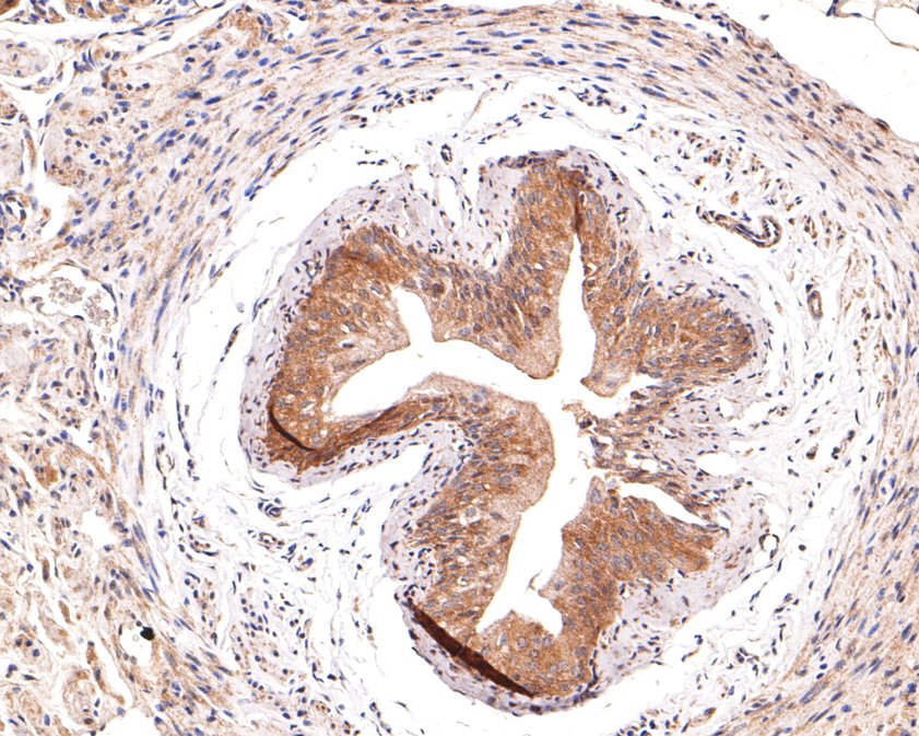
Immunohistochemical analysis of paraffin-embedded mouse bladder tissue using anti-IFNGR2 antibody. The section was pre-treated using heat mediated antigen retrieval with Tris-EDTA buffer (pH 9.0) for 20 minutes.The tissues were blocked in 1% BSA for 30 minutes at room temperature, washed with ddH2O and PBS, and then probed with the primary antibody (1/600) for 30 minutes at room temperature. The detection was performed using an HRP conjugated compact polymer system. DAB was used as the chromogen. Tissues were counterstained with hematoxylin and mounted with DPX.IHC
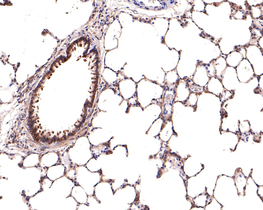
Immunohistochemical analysis of paraffin-embedded rat lung tissue using anti-IFNGR2 antibody. The section was pre-treated using heat mediated antigen retrieval with Tris-EDTA buffer (pH 9.0) for 20 minutes.The tissues were blocked in 1% BSA for 30 minutes at room temperature, washed with ddH2O and PBS, and then probed with the primary antibody (1/600) for 30 minutes at room temperature. The detection was performed using an HRP conjugated compact polymer system. DAB was used as the chromogen. Tissues were counterstained with hematoxylin and mounted with DPX.ICC/IF
ICC staining of IFNGR2 in SH-SY5Y cells (green). Formalin fixed cells were permeabilized with 0.1% Triton X-100 in TBS for 10 minutes at room temperature and blocked with 10% negative goat serum for 15 minutes at room temperature. Cells were probed with the primary antibody (1/200) for 1 hour at room temperature, washed with PBS. Alexa Fluor®488 conjugate-Goat anti-Rabbit IgG was used as the secondary antibody at 1/1,000 dilution. The nuclear counter stain is DAPI (blue).IF-P
Species: Mouse, Site: bladder, Sample: Paraffin-embedded section, Antibody concentration: 1/200.| Product Name | IFNGR2 Rabbit Polyclonal Antibody |
|---|---|
| Antibody Type | Primary Antibodies |
| Immunogen | Synthetic peptide within human IFN gamma Receptor beta aa 288-337. |
| Clonality | Polyclonal |
|---|---|
| Isotype | IgG |
| Host Species | Rabbit |
| Tested Applications | ICC/IFIF-PIHCWB |
| WB:1:1000 IHC:1:600 ICC/IF:1:200 IF-P:1:200 |
|
| Species Reactivity | HumanMouseRat |
| Concentration | 1mg/ml |
| Purification | Protein A |
| Gene Symbol | IFNGR2 |
|---|---|
| Gene Synonyms | AF-1 IFGR2 IMD28 IFNGT1 |
| Gene Full Name | interferon gamma receptor 2 |
| Gene Summary | This gene (IFNGR2) encodes the non-ligand-binding beta chain of the gamma interferon receptor. Human interferon-gamma receptor is a heterodimer of IFNGR1 and IFNGR2. Defects in IFNGR2 are a cause of mendelian susceptibility to mycobacterial disease (MSMD), also known as familial disseminated atypical mycobacterial infection. MSMD is a genetically heterogeneous disease with autosomal recessive, autosomal dominant or X-linked inheritance. [provided by RefSeq, Jul 2008] |
| Alternative Names | AF 1 antibody AF-1 antibody AF1 antibody IFGR 2 antibody IFGR2 antibody IFN gamma Receptor beta antibody IFN-gamma receptor 2 antibody IFN-gamma-R2 antibody IFNGR 2 antibody IFNGR2 antibody AF 1 antibody AF-1 antibody AF1 antibody IFGR 2 antibody IFGR2 antibody IFN gamma Receptor beta antibody IFN-gamma receptor 2 antibody IFN-gamma-R2 antibody IFNGR 2 antibody IFNGR2 antibody IFNGT 1 antibody IFNGT1 antibody IMD28 antibody INGR2_HUMAN antibody Interferon gamma receptor 2 (interferon gamma transducer 1) antibody Interferon gamma receptor 2 antibody Interferon gamma receptor accessory factor 1 antibody Interferon gamma receptor beta chain antibody Interferon gamma receptor chain 2 antibody Interferon gamma transducer 1 antibody |
| Molecular Weight(MW) | 38 kDa(Observed band size: 45 kDa) |
| Cellular Localization | Golgi apparatus membrane, Cell membrane, Cytoplasm, Endoplasmic reticulum membrane, Cytoplasmic vesicle membrane. |
WB
Western blot analysis of IFNGR2 on different lysates with Rabbit anti-IFNGR2 antibody at 1/1,000 dilution. Lane 1: HeLa cell lysate (20 µg/Lane), Lane 2: C2C12 cell lysate (20 µg/Lane), Lane 3: F9 cell lysate (20 µg/Lane), Lane 4: Mouse lung tissue lysate (40 µg/Lane), Lane 5: Mouse spleen tissue lysate (40 µg/Lane), Lane 6: Rat lung tissue lysate (40 µg/Lane), Lane 7: Rat spleen tissue lysate (40 µg/Lane), Exposure time: 1 minute; 4-20% SDS-PAGE gel. Proteins were transferred to a PVDF membrane and blocked with 5% NFDM/TBST for 1 hour at room temperature. The primary antibody at 1/1,000 dilution was used in 5% NFDM/TBST at 4℃ overnight. Goat Anti-Rabbit IgG - HRP Secondary Antibody at 1/50,000 dilution was used for 1 hour at room temperature.
IHC
Immunohistochemical analysis of paraffin-embedded human stomach tissue using anti-IFNGR2 antibody. The section was pre-treated using heat mediated antigen retrieval with Tris-EDTA buffer (pH 9.0) for 20 minutes.The tissues were blocked in 1% BSA for 30 minutes at room temperature, washed with ddH2O and PBS, and then probed with the primary antibody (1/600) for 30 minutes at room temperature. The detection was performed using an HRP conjugated compact polymer system. DAB was used as the chromogen. Tissues were counterstained with hematoxylin and mounted with DPX.
IHC
Immunohistochemical analysis of paraffin-embedded mouse bladder tissue using anti-IFNGR2 antibody. The section was pre-treated using heat mediated antigen retrieval with Tris-EDTA buffer (pH 9.0) for 20 minutes.The tissues were blocked in 1% BSA for 30 minutes at room temperature, washed with ddH2O and PBS, and then probed with the primary antibody (1/600) for 30 minutes at room temperature. The detection was performed using an HRP conjugated compact polymer system. DAB was used as the chromogen. Tissues were counterstained with hematoxylin and mounted with DPX.
IHC
Immunohistochemical analysis of paraffin-embedded rat lung tissue using anti-IFNGR2 antibody. The section was pre-treated using heat mediated antigen retrieval with Tris-EDTA buffer (pH 9.0) for 20 minutes.The tissues were blocked in 1% BSA for 30 minutes at room temperature, washed with ddH2O and PBS, and then probed with the primary antibody (1/600) for 30 minutes at room temperature. The detection was performed using an HRP conjugated compact polymer system. DAB was used as the chromogen. Tissues were counterstained with hematoxylin and mounted with DPX.
ICC/IF
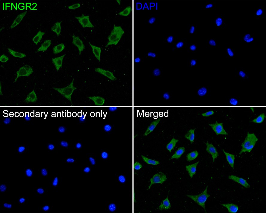
ICC staining of IFNGR2 in SH-SY5Y cells (green). Formalin fixed cells were permeabilized with 0.1% Triton X-100 in TBS for 10 minutes at room temperature and blocked with 10% negative goat serum for 15 minutes at room temperature. Cells were probed with the primary antibody (1/200) for 1 hour at room temperature, washed with PBS. Alexa Fluor®488 conjugate-Goat anti-Rabbit IgG was used as the secondary antibody at 1/1,000 dilution. The nuclear counter stain is DAPI (blue).
IF-P
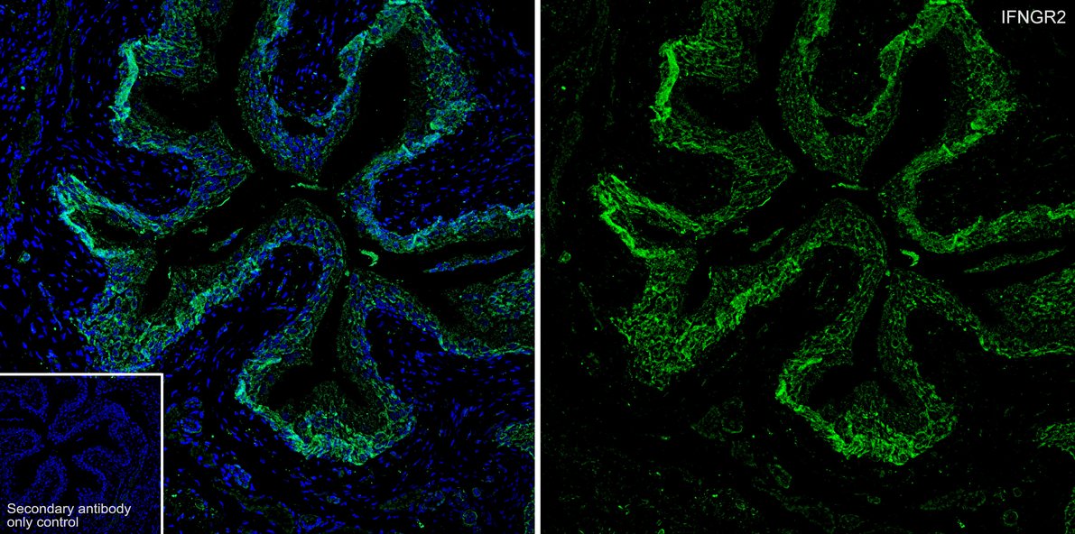
Species: Mouse, Site: bladder, Sample: Paraffin-embedded section, Antibody concentration: 1/200.| Application Notes | WB:1:1000 IHC:1:600 ICC/IF:1:200 IF-P:1:200 |
|---|
| Form | Liquid |
|---|---|
| Storage Instructions | Shipped at 4℃. Store at +4℃ short term (1-2 weeks). It is recommended to aliquot into single-use upon delivery. Store at -20℃ long term. |
| Storage Buffer | PBS (pH7.4), 0.1% BSA, 40% Glycerol. Preservative: 0.05% Sodium Azide. |


