WB
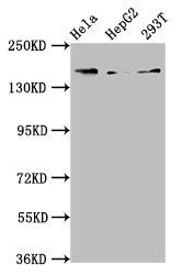
Positive WB detected in: Hela whole cell lysate, HepG2 whole cell lysate, 293T whole cell lysate, All lanes: C4A antibody at 3.9μg/ml. Secondary Goat polyclonal to rabbit IgG at 1/50000 dilution.IHC
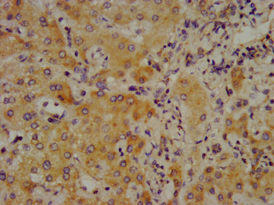
IHC image of C4A Antibody diluted at 1:700 and staining in paraffin-embedded human liver cancer performed on a Leica BondTM system. After dewaxing and hydration, antigen retrieval was mediated by high pressure in a citrate buffer (pH 6.0). Section was blocked with 10% normal goat serum 30min at RT. Then primary antibody (1% BSA) was incubated at 4°C overnight. The primary is detected by a biotinylated secondary antibody and visualized using an HRP conjugated SP system.| Product Name | Rabbit anti-C4A Polyclonal antibody |
|---|---|
| Antibody Type | Primary Antibodies |
| Immunogen | Recombinant Human Complement C4-A protein (1027-1186AA). |
| Clonality | Polyclonal |
|---|---|
| Isotype | IgG |
| Host Species | Rabbit |
| Tested Applications | ICC/IFIHCWB |
| WB:1:500-1:5000 IHC:1:500-1:1000 ICC/IF:1:50-1:200 |
|
| Species Reactivity | Human |
| Concentration | 1mg/ml |
| Purification | Affinity purified |
| Gene Symbol | C4A |
|---|---|
| Gene Synonyms | C4 RG C4S CO4 C4A2 C4A3 C4A4 C4A6 C4AD CPAMD2 |
| Gene Full Name | complement C4A |
| Gene Summary | This gene encodes the acidic form of complement factor 4, part of the classical activation pathway. The protein is expressed as a single chain precursor which is proteolytically cleaved into a trimer of alpha, beta, and gamma chains prior to secretion. The trimer provides a surface for interaction between the antigen-antibody complex and other complement components. The alpha chain is cleaved to release C4 anaphylatoxin, an antimicrobial peptide and a mediator of local inflammation. Deficiency of this protein is associated with systemic lupus erythematosus and type I diabetes mellitus. This gene localizes to the major histocompatibility complex (MHC) class III region on chromosome 6. Varying haplotypes of this gene cluster exist, such that individuals may have 1, 2, or 3 copies of this gene. Two transcript variants encoding different isoforms have been found for this gene. [provided by RefSeq, Nov 2014] |
| Alternative Names | Acidic C4 antibody Acidic complement C4 antibody Basic C4 antibody Basic complement C4 antibody C3 and PZP-like alpha-2-macroglobulin domain-containing protein 2 antibody C3 and PZP-like alpha-2-macroglobulin domain-containing protein 3 antibody C4 Chido form antibody C4 Rodgers from antibody C4A anaphylatoxin antibody C4A antibody C4A2 antibody C4A3 antibody C4A4 antibody C4A6 antibody C4AD antibody C4B antibody C4B_2 antibody C4B1 antibody C4B12 antibody C4B2 antibody C4B3 antibody C4BD antibody C4F antibody C4S antibody CH antibody Chido form of C4 antibody CO4 antibody CO4A_HUMAN antibody Complement C4 A antibody Complement C4 B antibody Complement C4 gamma chain antibody complement C4-A antibody complement C4-B antibody complement C4-B-like antibody complement C4B1a antibody Complement component 4A (Rodgers blood group) antibody Complement component 4A antibody complement component 4B (Chido blood group) antibody complement component 4B (Chido blood group) copy 2 antibody Complement component 4B (Childo blood group) antibody Complement component 4B antibody Complement component 4F antibody Complement component 4S antibody CPAMD2 antibody CPAMD3 antibody RG antibody Rodgers form of C4 antibody |
| Molecular Weight(MW) | 193 kDa |
| Cellular Localization | Secreted. Cell junction, synapse. Cell projection, axon. Cell projection, dendrite. |
WB
Positive WB detected in: Hela whole cell lysate, HepG2 whole cell lysate, 293T whole cell lysate, All lanes: C4A antibody at 3.9μg/ml. Secondary Goat polyclonal to rabbit IgG at 1/50000 dilution.
IHC
IHC image of C4A Antibody diluted at 1:700 and staining in paraffin-embedded human liver cancer performed on a Leica BondTM system. After dewaxing and hydration, antigen retrieval was mediated by high pressure in a citrate buffer (pH 6.0). Section was blocked with 10% normal goat serum 30min at RT. Then primary antibody (1% BSA) was incubated at 4°C overnight. The primary is detected by a biotinylated secondary antibody and visualized using an HRP conjugated SP system.| Application Notes | WB:1:500-1:5000 IHC:1:500-1:1000 ICC/IF:1:50-1:200 |
|---|
| Form | Liquid |
|---|---|
| Storage Instructions | Upon receipt, store at -20°C or -80°C. Avoid repeated freeze. |
| Storage Buffer | 0.03% Proclin 300 Constituents: 50% Glycerol, 0.01M PBS, PH 7.4 |


