WB
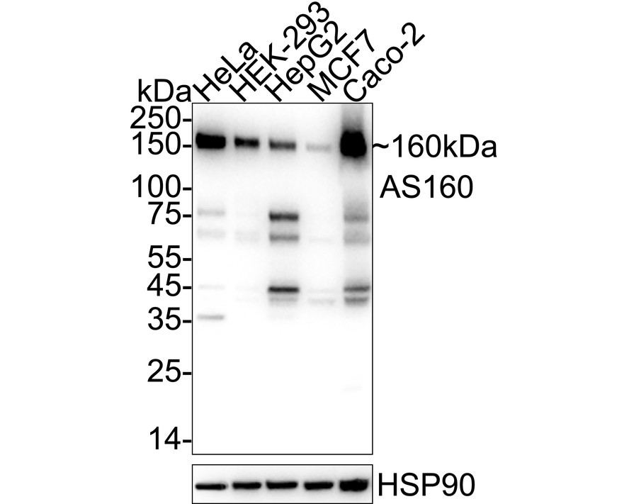
Western blot analysis of AS160 on different lysates with Rabbit anti-AS160 antibody at 1/2,000 dilution. Exposure time: 10 seconds; 4-20% SDS-PAGE gel. Proteins were transferred to a PVDF membrane and blocked with 5% NFDM/TBST for 1 hour at room temperature. The primary antibody at 1/2,000 dilution was used in 5% NFDM/TBST at 4℃ overnight. Goat Anti-Rabbit IgG - HRP Secondary Antibody at 1/50,000 dilution was used for 1 hour at room temperature.IHC
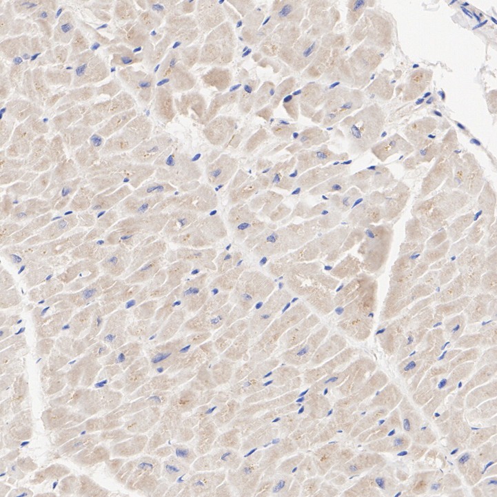
Immunohistochemical analysis of paraffin-embedded human heart tissue with Rabbit anti-AS160 antibody at 1/1,000 dilution. The section was pre-treated using heat mediated antigen retrieval with Tris-EDTA buffer (pH 9.0) for 20 minutes. The tissues were blocked in 1% BSA for 20 minutes at room temperature, washed with ddH2O and PBS, and then probed with the primary antibody at 1/1,000 dilution for 1 hour at room temperature. The detection was performed using an HRP conjugated compact polymer system. DAB was used as the chromogen. Tissues were counterstained with hematoxylin and mounted with DPX.IHC
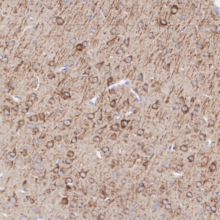
Immunohistochemical analysis of paraffin-embedded rat brain tissue with Rabbit anti-AS160 antibody at 1/1,000 dilution. The section was pre-treated using heat mediated antigen retrieval with Tris-EDTA buffer (pH 9.0) for 20 minutes. The tissues were blocked in 1% BSA for 20 minutes at room temperature, washed with ddH2O and PBS, and then probed with the primary antibody at 1/1,000 dilution for 1 hour at room temperature. The detection was performed using an HRP conjugated compact polymer system. DAB was used as the chromogen. Tissues were counterstained with hematoxylin and mounted with DPX.IP
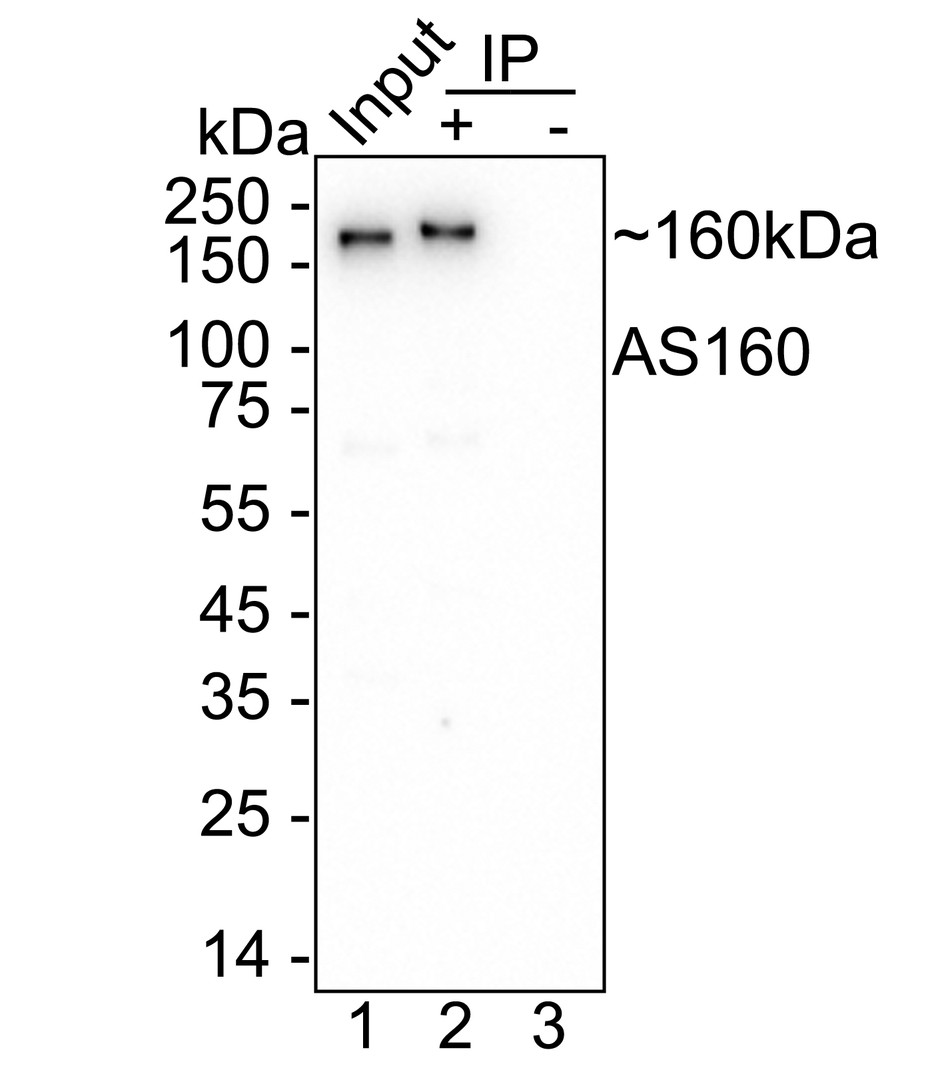
AS160 was immunoprecipitated from 0.2 mg HeLa cell lysate with AS160 Recombinant Rabbit Monoclonal Antibodyat 2 µg/10 µl beads. Western blot was performed from the immunoprecipitate usingAS160 Recombinant Rabbit Monoclonal Antibodyat 1/1,000 dilution. HRP Conjugated Anti-Rabbit IgG for IP Nano-secondary antibody at 1/5,000 dilution was used for 1 hour at room temperature. Lane 1: HeLa cell lysate (input) Lane 2: AS160 Recombinant Rabbit Monoclonal Antibody IP in HeLa cell lysate Lane 3: Rabbit IgG instead of AS160 Recombinant Rabbit Monoclonal Antibodyin HeLa cell lysate Blocking/Dilution buffer: 5% NFDM/TBST Exposure time: 9 seconds;| Product Name | AS160 Recombinant Rabbit Monoclonal Antibody |
|---|---|
| Antibody Type | Primary Antibodies |
| Immunogen | Recombinant protein within human AS160 aa 1-500. |
| Clonality | Monoclonal |
|---|---|
| Isotype | IgG |
| Host Species | Rabbit |
| Tested Applications | IHCIPWB |
| WB:1:2000-1:10000 IHC:1:200-1:1000 WB:1-2μg/sample IP:1:10-1:100 |
|
| Species Reactivity | HumanRat |
| Concentration | 1mg/ml |
| Purification | Protein A |
| Gene Symbol | TBC1D4 |
|---|---|
| Gene Synonyms | AS160 NIDDM5 |
| Gene Full Name | TBC1 domain family member 4 |
| Gene Summary | This gene is a member of the Tre-2/BUB2/CDC16 domain family. The protein encoded by this gene is a Rab-GTPase-activating protein, and contains two phopshotyrosine-binding domains (PTB1 and PTB2), a calmodulin-binding domain (CBD), a Rab-GTPase domain, and multiple AKT phosphomotifs. This protein is thought to play an important role in glucose homeostasis by regulating the insulin-dependent trafficking of the glucose transporter 4 (GLUT4), important for removing glucose from the bloodstream into skeletal muscle and fat tissues. Reduced expression of this gene results in an increase in GLUT4 levels at the plasma membrane, suggesting that this protein is important in intracellular retention of GLUT4 under basal conditions. When exposed to insulin, this protein is phosphorylated, dissociates from GLUT4 vesicles, resulting in increased GLUT4 at the cell surface, and enhanced glucose transport. Phosphorylation of this protein by AKT is required for proper translocation of GLUT4 to the cell surface. Individuals homozygous for a mutation in this gene are at higher risk for type 2 diabetes and have higher levels of circulating glucose and insulin levels after glucose ingestion. Alternative splicing results in multiple transcript variants encoding different isoforms. [provided by RefSeq, Aug 2015] |
| Alternative Names | Acrg embryonic lethality (mouse) minimal region ortholog antibody Acrg embryonic lethality minimal region ortholog antibody Acrg embryonic lethality mouse minimal region ortholog antibody Akt substrate of 160 kDa antibody AS 160 antibody AS160 antibody BUB2 antibody CDC16 antibody KIAA0603 antibody NIDDM5 antibody Acrg embryonic lethality (mouse) minimal region ortholog antibody Acrg embryonic lethality minimal region ortholog antibody Acrg embryonic lethality mouse minimal region ortholog antibody Akt substrate of 160 kDa antibody AS 160 antibody AS160 antibody BUB2 antibody CDC16 antibody KIAA0603 antibody NIDDM5 antibody TBC (Tre 2 BUB2 CDC16) domain containing protein antibody TBC Tre 2 BUB2 CDC16 domain containing protein antibody TBC1 D4 antibody TBC1 domain family member 4 antibody Tbc1d4 antibody TBCD4_HUMAN antibody Tre-2 antibody |
| Molecular Weight(MW) | 147 kDa(Observed band size: 160 kDa) |
| Cellular Localization | Cytoplasm. |
WB
Western blot analysis of AS160 on different lysates with Rabbit anti-AS160 antibody at 1/2,000 dilution. Exposure time: 10 seconds; 4-20% SDS-PAGE gel. Proteins were transferred to a PVDF membrane and blocked with 5% NFDM/TBST for 1 hour at room temperature. The primary antibody at 1/2,000 dilution was used in 5% NFDM/TBST at 4℃ overnight. Goat Anti-Rabbit IgG - HRP Secondary Antibody at 1/50,000 dilution was used for 1 hour at room temperature.
IHC
Immunohistochemical analysis of paraffin-embedded human heart tissue with Rabbit anti-AS160 antibody at 1/1,000 dilution. The section was pre-treated using heat mediated antigen retrieval with Tris-EDTA buffer (pH 9.0) for 20 minutes. The tissues were blocked in 1% BSA for 20 minutes at room temperature, washed with ddH2O and PBS, and then probed with the primary antibody at 1/1,000 dilution for 1 hour at room temperature. The detection was performed using an HRP conjugated compact polymer system. DAB was used as the chromogen. Tissues were counterstained with hematoxylin and mounted with DPX.
IHC
Immunohistochemical analysis of paraffin-embedded rat brain tissue with Rabbit anti-AS160 antibody at 1/1,000 dilution. The section was pre-treated using heat mediated antigen retrieval with Tris-EDTA buffer (pH 9.0) for 20 minutes. The tissues were blocked in 1% BSA for 20 minutes at room temperature, washed with ddH2O and PBS, and then probed with the primary antibody at 1/1,000 dilution for 1 hour at room temperature. The detection was performed using an HRP conjugated compact polymer system. DAB was used as the chromogen. Tissues were counterstained with hematoxylin and mounted with DPX.
IP
AS160 was immunoprecipitated from 0.2 mg HeLa cell lysate with AS160 Recombinant Rabbit Monoclonal Antibodyat 2 µg/10 µl beads. Western blot was performed from the immunoprecipitate usingAS160 Recombinant Rabbit Monoclonal Antibodyat 1/1,000 dilution. HRP Conjugated Anti-Rabbit IgG for IP Nano-secondary antibody at 1/5,000 dilution was used for 1 hour at room temperature. Lane 1: HeLa cell lysate (input) Lane 2: AS160 Recombinant Rabbit Monoclonal Antibody IP in HeLa cell lysate Lane 3: Rabbit IgG instead of AS160 Recombinant Rabbit Monoclonal Antibodyin HeLa cell lysate Blocking/Dilution buffer: 5% NFDM/TBST Exposure time: 9 seconds;| Application Notes | WB:1:2000-1:10000 IHC:1:200-1:1000 WB:1-2μg/sample IP:1:10-1:100 |
|---|
| Form | Liquid |
|---|---|
| Storage Instructions | Shipped at 4℃. Store at +4℃ short term (1-2 weeks). It is recommended to aliquot into single-use upon delivery. Store at -20℃ long term. |
| Storage Buffer | PBS (pH7.4), 0.1% BSA, 40% Glycerol. Preservative: 0.05% Sodium Azide. |


