WB
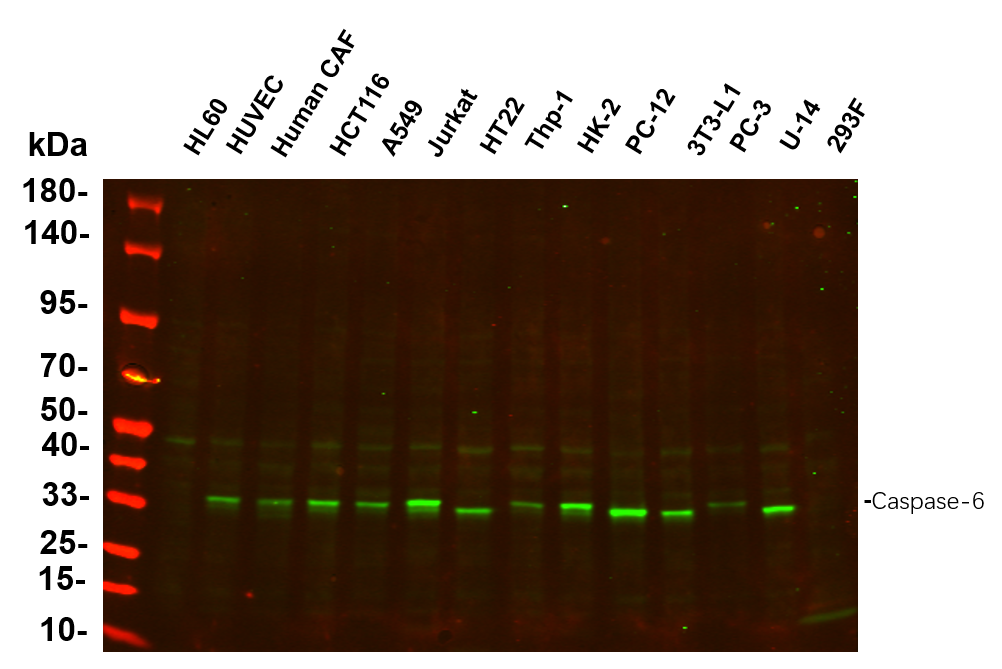
Various whole cell lysates were separated by 4-20% SDS-PAGE, and the primary antibody was used at 4℃, over night with a 1:5000 dilution . The Dylight 800-conjugated Goat anti-Rabbit antibody was used to detect the antibody.WB
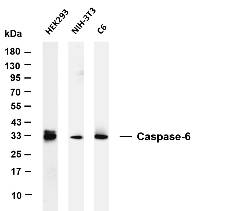
Various whole cell lysates were separated by 4-20% SDS-PAGE, and the membrane was blotted with anti-Caspase-6 antibody. The HRP-conjugated Goat anti-Rabbit IgG(H + L) antibody was used to detect the antibody.IHC
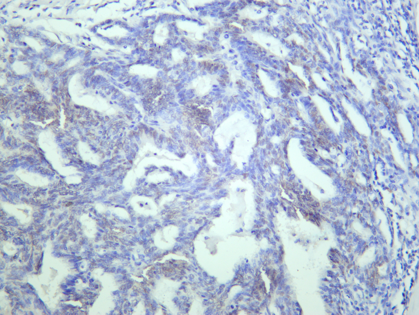
Human endometrium carcinoma was stained with anti-Caspase-6 Rabbit antibody.IHC
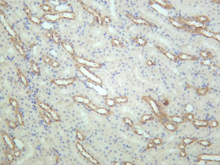
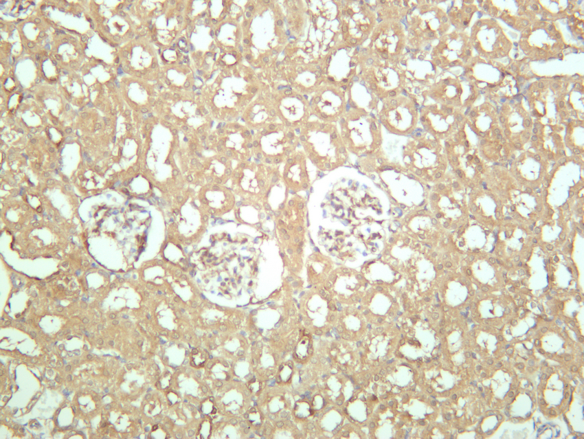
Mouse kidney was stained with anti-Caspase-6 Rabbit antibody.IHC
Rat kidney was stained with anti-Caspase-6 Rabbit antibody.ICC/IF
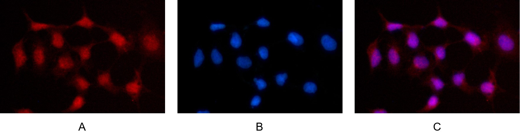
Immunofluorescence analysis of HEK293. Picture A: Caspase-6 antibody (red). Picture B: DAPI (blue). Picture C: Merge of A+B.| Product Name | Caspase-6 Rabbit mAb |
|---|---|
| Antibody Type | Primary Antibodies |
| Clonality | Monoclonal |
|---|---|
| Isotype | IgG,K |
| Host Species | Rabbit |
| Tested Applications | ICC/IFIHCWB |
| WB:1:2000-1:10000 IHC:1:200-1:500 ICC/IF:1:200-1:1000 |
|
| Species Reactivity | HumanMouseRat |
| Concentration | 1mg/ml |
| Purification | Protein A |
| Gene Symbol | CASP6 |
|---|---|
| Gene Synonyms | MCH2 CSP-6 caspase-6 |
| Gene Full Name | caspase 6 |
| Gene Summary | This gene encodes a member of the cysteine-aspartic acid protease (caspase) family of enzymes. Sequential activation of caspases plays a central role in the execution-phase of cell apoptosis. Caspases exist as inactive proenzymes which undergo proteolytic processing at conserved aspartic acid residues to produce two subunits, large and small, that dimerize to form the active enzyme. This protein is processed by caspases 7, 8 and 10, and is thought to function as a downstream enzyme in the caspase activation cascade. Alternative splicing of this gene results in multiple transcript variants that encode different isoforms. [provided by RefSeq, Oct 2015] |
| Alternative Names | CASP6 MCH2 Caspase-6 CASP-6 Apoptotic protease Mch-2 |
| Molecular Weight(MW) | 33 kDa |
| Cellular Localization | Cytoplasm. Nucleus. |
WB
Various whole cell lysates were separated by 4-20% SDS-PAGE, and the primary antibody was used at 4℃, over night with a 1:5000 dilution . The Dylight 800-conjugated Goat anti-Rabbit antibody was used to detect the antibody.
WB
Various whole cell lysates were separated by 4-20% SDS-PAGE, and the membrane was blotted with anti-Caspase-6 antibody. The HRP-conjugated Goat anti-Rabbit IgG(H + L) antibody was used to detect the antibody.
IHC
Human endometrium carcinoma was stained with anti-Caspase-6 Rabbit antibody.
IHC
Mouse kidney was stained with anti-Caspase-6 Rabbit antibody.
IHC
Rat kidney was stained with anti-Caspase-6 Rabbit antibody.
ICC/IF
Immunofluorescence analysis of HEK293. Picture A: Caspase-6 antibody (red). Picture B: DAPI (blue). Picture C: Merge of A+B.| Application Notes | WB:1:2000-1:10000 IHC:1:200-1:500 ICC/IF:1:200-1:1000 |
|---|
| Form | Liquid |
|---|---|
| Storage Instructions | -15°C to -25°C/1 year(Do not lower than -25°C) |
| Storage Buffer | PBS, 50% glycerol, 0.05% Proclin 300, 0.05%BSA |


