WB
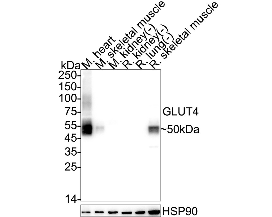
Western blot analysis of Glucose Transporter GLUT4 on different lysates with Rabbit anti-Glucose Transporter GLUT4 antibody at 1/5,000 dilution. Lane 1: Mouse heart tissue lysate (no heat) (30 µg/Lane), Lane 2: Mouse skeletal muscle tissue lysate (no heat) (30 µg/Lane), Lane 3: Mouse kidney tissue lysate (no heat) (negative) (30 µg/Lane), Lane 4: Rat kidney tissue lysate (negative) (30 µg/Lane), Lane 5: Rat lung tissue lysate (no heat) (negative) (30 µg/Lane), Lane 6: Rat skeletal muscle tissue lysate (no heat) (30 µg/Lane), Notice: no heat means the lysate is not boiled. Exposure time: 20 seconds; 4-20% SDS-PAGE gel. Proteins were transferred to a PVDF membrane and blocked with 5% NFDM/TBST for 1 hour at room temperature. The primary antibody at 1/5,000 dilution was used in 5% NFDM/TBST at 4℃ overnight. Goat Anti-Rabbit IgG - HRP Secondary Antibody at 1/50,000 dilution was used for 1 hour at room temperature.IF-F
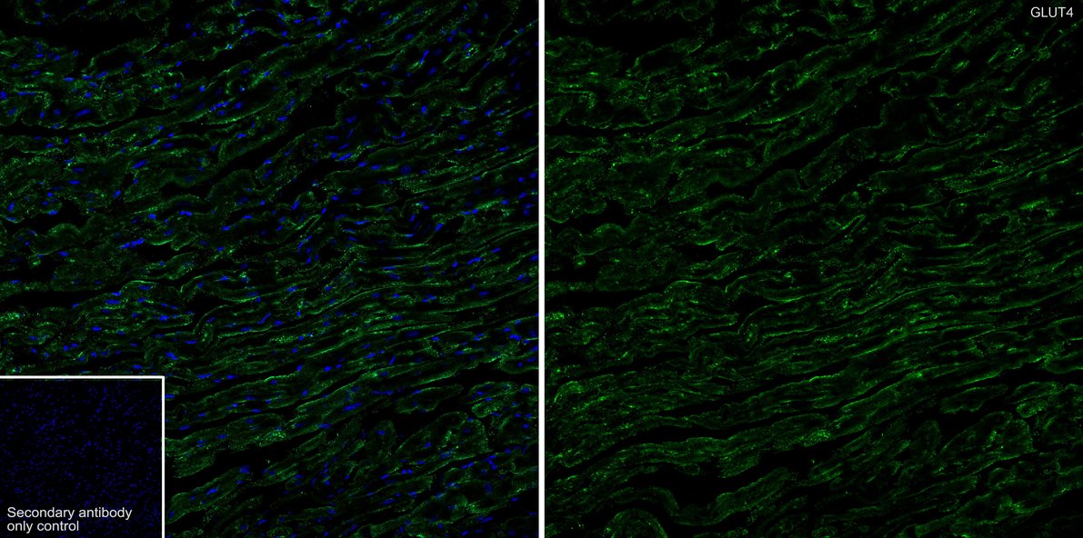
Immunofluorescence analysis of frozen mouse heart tissue with Rabbit anti-Glucose Transporter GLUT4 antibody at 1/500 dilution. The section was not undergone antigen retrieval. The tissues were blocked in 10% negative goat serum for 1 hour at room temperature, washed with PBS, and then probed with the primary antibody ( green) at 1/500 dilution overnight at 4 ℃, washed with PBS. Goat Anti-Rabbit IgG H&L (488) was used as the secondary antibody at 1/1,000 dilution. Nuclei were counterstained with DAPI (blue).IF-F
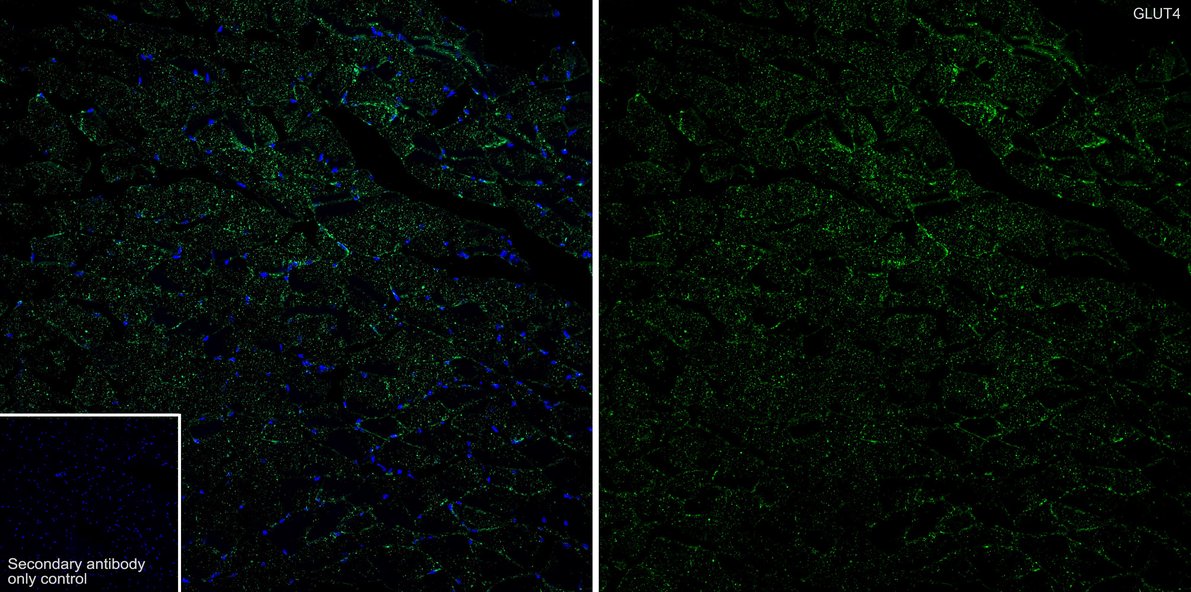
Immunofluorescence analysis of frozen rat skeletal muscle tissue with Rabbit anti-Glucose Transporter GLUT4 antibody at 1/500 dilution. The section was not undergone antigen retrieval. The tissues were blocked in 10% negative goat serum for 1 hour at room temperature, washed with PBS, and then probed with the primary antibody (green) at 1/500 dilution overnight at 4 ℃, washed with PBS. Goat Anti-Rabbit IgG H&L (488) was used as the secondary antibody at 1/1,000 dilution. Nuclei were counterstained with DAPI (blue).| Product Name | Glucose Transporter GLUT4 Recombinant Rabbit Monoclonal Antibody |
|---|---|
| Antibody Type | Primary Antibodies |
| Immunogen | Synthetic peptide within human GLUT4 aa 447-509. |
| Clonality | Monoclonal |
|---|---|
| Isotype | IgG |
| Host Species | Rabbit |
| Tested Applications | IF-FWB |
| WB:1:5000 IF-F:1:500 |
|
| Species Reactivity | MouseRat |
| Concentration | 1mg/ml |
| Purification | Protein A |
| Gene Symbol | SLC2A4 |
|---|---|
| Gene Synonyms | GLUT4 |
| Gene Full Name | solute carrier family 2 member 4 |
| Gene Summary | This gene is a member of the solute carrier family 2 (facilitated glucose transporter) family and encodes a protein that functions as an insulin-regulated facilitative glucose transporter. In the absence of insulin, this integral membrane protein is sequestered within the cells of muscle and adipose tissue. Within minutes of insulin stimulation, the protein moves to the cell surface and begins to transport glucose across the cell membrane. Mutations in this gene have been associated with noninsulin-dependent diabetes mellitus (NIDDM). [provided by RefSeq, Jul 2008] |
| Alternative Names | insulin-responsive antibody Glucose transporter GLUT 4 antibody Glucose transporter type 4 antibody Glucose transporter type 4 insulin responsive antibody GLUT 4 antibody GLUT-4 antibody GLUT4 antibody GTR4_HUMAN antibody Insulin responsive glucose transporter type 4 antibody kug antibody insulin-responsive antibody Glucose transporter GLUT 4 antibody Glucose transporter type 4 antibody Glucose transporter type 4 insulin responsive antibody GLUT 4 antibody GLUT-4 antibody GLUT4 antibody GTR4_HUMAN antibody Insulin responsive glucose transporter type 4 antibody kug antibody SLC 2A4 antibody SLC2A4 antibody solute carrier family 2 (facilitated glucose transporter) member 4 antibody Solute carrier family 2 member 4 antibody Solute carrier family 2 facilitated glucose transporter member 4 antibody |
| Molecular Weight(MW) | 55 kDa(Observed band size: 50 kDa) |
| Cellular Localization | Cell membrane, Endomembrane system, Cytoplasm, perinuclear region. |
WB
Western blot analysis of Glucose Transporter GLUT4 on different lysates with Rabbit anti-Glucose Transporter GLUT4 antibody at 1/5,000 dilution. Lane 1: Mouse heart tissue lysate (no heat) (30 µg/Lane), Lane 2: Mouse skeletal muscle tissue lysate (no heat) (30 µg/Lane), Lane 3: Mouse kidney tissue lysate (no heat) (negative) (30 µg/Lane), Lane 4: Rat kidney tissue lysate (negative) (30 µg/Lane), Lane 5: Rat lung tissue lysate (no heat) (negative) (30 µg/Lane), Lane 6: Rat skeletal muscle tissue lysate (no heat) (30 µg/Lane), Notice: no heat means the lysate is not boiled. Exposure time: 20 seconds; 4-20% SDS-PAGE gel. Proteins were transferred to a PVDF membrane and blocked with 5% NFDM/TBST for 1 hour at room temperature. The primary antibody at 1/5,000 dilution was used in 5% NFDM/TBST at 4℃ overnight. Goat Anti-Rabbit IgG - HRP Secondary Antibody at 1/50,000 dilution was used for 1 hour at room temperature.
IF-F
Immunofluorescence analysis of frozen mouse heart tissue with Rabbit anti-Glucose Transporter GLUT4 antibody at 1/500 dilution. The section was not undergone antigen retrieval. The tissues were blocked in 10% negative goat serum for 1 hour at room temperature, washed with PBS, and then probed with the primary antibody ( green) at 1/500 dilution overnight at 4 ℃, washed with PBS. Goat Anti-Rabbit IgG H&L (488) was used as the secondary antibody at 1/1,000 dilution. Nuclei were counterstained with DAPI (blue).
IF-F
Immunofluorescence analysis of frozen rat skeletal muscle tissue with Rabbit anti-Glucose Transporter GLUT4 antibody at 1/500 dilution. The section was not undergone antigen retrieval. The tissues were blocked in 10% negative goat serum for 1 hour at room temperature, washed with PBS, and then probed with the primary antibody (green) at 1/500 dilution overnight at 4 ℃, washed with PBS. Goat Anti-Rabbit IgG H&L (488) was used as the secondary antibody at 1/1,000 dilution. Nuclei were counterstained with DAPI (blue).| Application Notes | WB:1:5000 IF-F:1:500 |
|---|
| Form | Liquid |
|---|---|
| Storage Instructions | Shipped at 4℃. Store at +4℃ short term (1-2 weeks). It is recommended to aliquot into single-use upon delivery. Store at -20℃ long term. |
| Storage Buffer | PBS (pH7.4), 0.1% BSA, 40% Glycerol. Preservative: 0.05% Sodium Azide. |


