WB
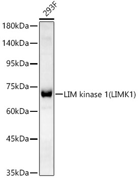
Western blot analysis of lysates from 293F cells, using LIM kinase 1(LIMK1) Rabbit mAb at 1:2000 dilution. Secondary antibody: HRP-conjugated Goat anti-Rabbit IgG (H+L) at 1:10000 dilution. Lysates/proteins: 25μg per lane. Blocking buffer: 3% nonfat dry milk in TBST. Detection: ECL Basic Kit. Exposure time: 90s.IHC
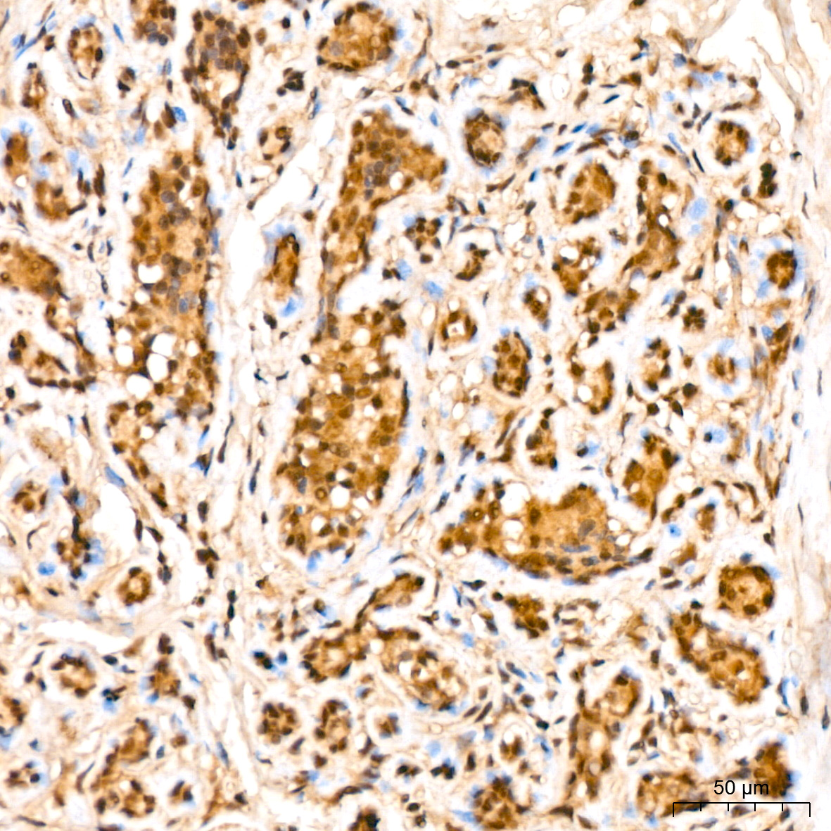
Immunohistochemistry analysis of paraffin embedded Human breast using LIM kinase 1(LIMK1) Rabbit mAb at dilution of 1:200 (40x lens). High pressure antigen retrieval performed with 0.01M Citrate buffer (pH 6.0) prior to IHC staining.IHC
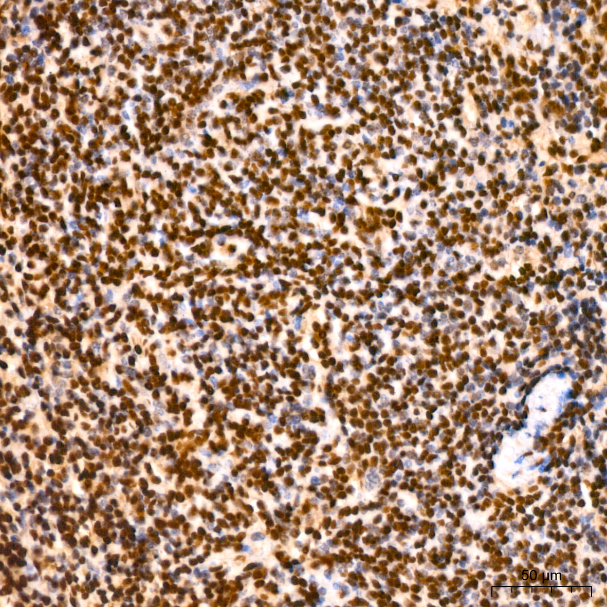
Immunohistochemistry analysis of paraffin embedded Mouse spleen using LIM kinase 1(LIMK1) Rabbit mAb at dilution of 1:200 (40x lens). High pressure antigen retrieval performed with 0.01M Citrate buffer (pH 6.0) prior to IHC staining.IHC
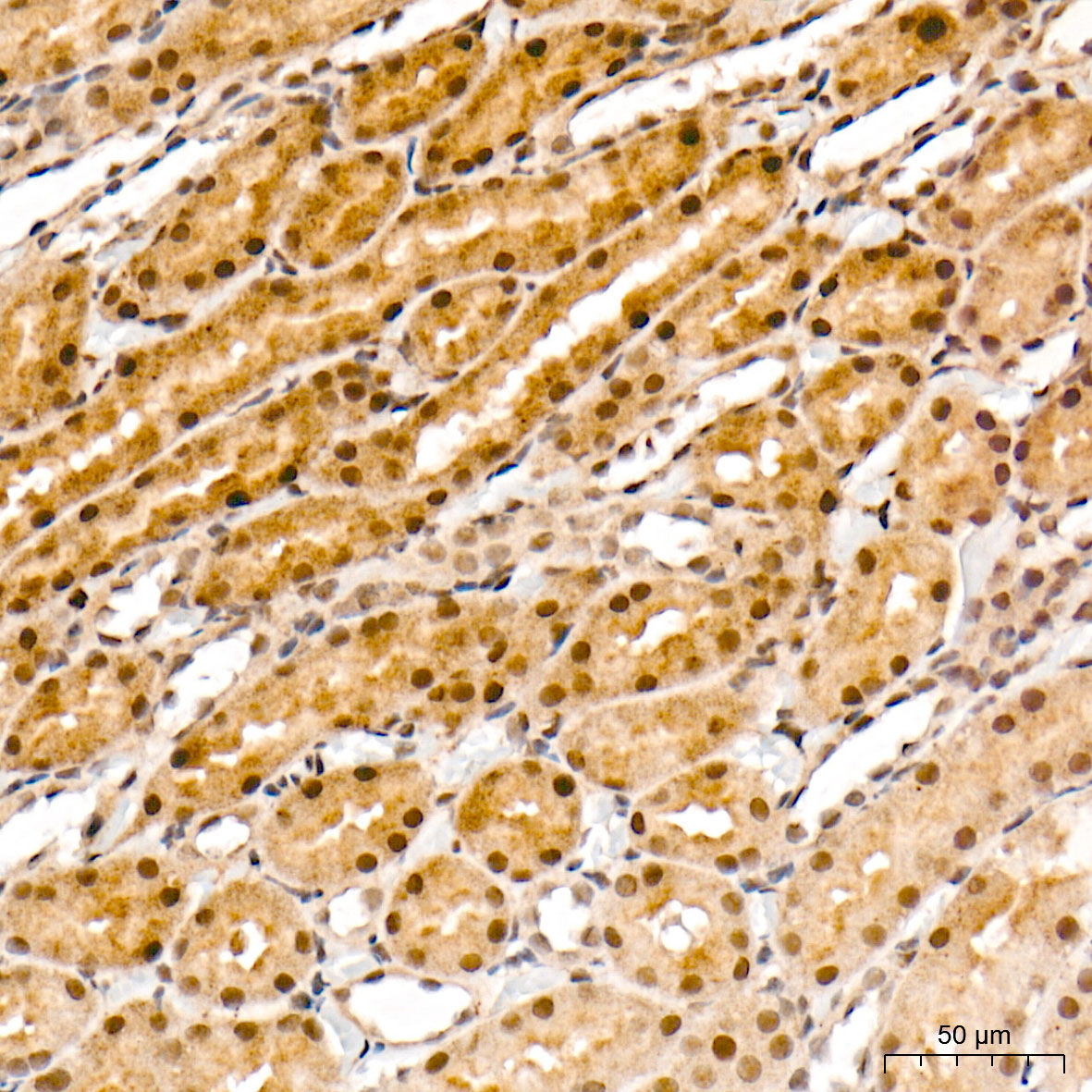
Immunohistochemistry analysis of paraffin embedded Rat kidney tissue using LIM kinase 1(LIMK1) Rabbit mAb at a dilution of 1:200 (40x lens). High pressure antigen retrieval performed with 0.01M Tris EDTA Buffer (pH 9.0) prior to IHC staining.| Product Name | LIM kinase 1(LIMK1) Rabbit mAb |
|---|---|
| Antibody Type | Primary Antibodies |
| Immunogen | Synthetic peptide. |
| Clonality | Monoclonal |
|---|---|
| Isotype | IgG |
| Host Species | Rabbit |
| Tested Applications | IHCWB |
| WB:1:1000-1:5000 IHC:1:50-1:200 |
|
| Species Reactivity | HumanMouseRat |
| Concentration | 1mg/ml |
| Purification | Affinity purified |
| Gene Symbol | LIMK1 |
|---|---|
| Gene Synonyms | LIMK LIMK-1 |
| Gene Full Name | LIM domain kinase 1 |
| Gene Summary | There are approximately 40 known eukaryotic LIM proteins, so named for the LIM domains they contain. LIM domains are highly conserved cysteine-rich structures containing 2 zinc fingers. Although zinc fingers usually function by binding to DNA or RNA, the LIM motif probably mediates protein-protein interactions. LIM kinase-1 and LIM kinase-2 belong to a small subfamily with a unique combination of 2 N-terminal LIM motifs and a C-terminal protein kinase domain. LIMK1 is a serine/threonine kinase that regulates actin polymerization via phosphorylation and inactivation of the actin binding factor cofilin. This protein is ubiquitously expressed during development and plays a role in many cellular processes associated with cytoskeletal structure. This protein also stimulates axon growth and may play a role in brain development. LIMK1 hemizygosity is implicated in the impaired visuospatial constructive cognition of Williams syndrome. Alternative splicing results in multiple transcript variants encoding distinct isoforms.[provided by RefSeq, Feb 2011] |
| Alternative Names | LIMK1 LIMK LIMK-1 LIM domain kinase 1 LIM kinase 1(LIMK1) |
| Molecular Weight(MW) | 72 kDa |
| Cellular Localization | Cytoplasm, Nucleus. |
WB
Western blot analysis of lysates from 293F cells, using LIM kinase 1(LIMK1) Rabbit mAb at 1:2000 dilution. Secondary antibody: HRP-conjugated Goat anti-Rabbit IgG (H+L) at 1:10000 dilution. Lysates/proteins: 25μg per lane. Blocking buffer: 3% nonfat dry milk in TBST. Detection: ECL Basic Kit. Exposure time: 90s.
IHC
Immunohistochemistry analysis of paraffin embedded Human breast using LIM kinase 1(LIMK1) Rabbit mAb at dilution of 1:200 (40x lens). High pressure antigen retrieval performed with 0.01M Citrate buffer (pH 6.0) prior to IHC staining.
IHC
Immunohistochemistry analysis of paraffin embedded Mouse spleen using LIM kinase 1(LIMK1) Rabbit mAb at dilution of 1:200 (40x lens). High pressure antigen retrieval performed with 0.01M Citrate buffer (pH 6.0) prior to IHC staining.
IHC
Immunohistochemistry analysis of paraffin embedded Rat kidney tissue using LIM kinase 1(LIMK1) Rabbit mAb at a dilution of 1:200 (40x lens). High pressure antigen retrieval performed with 0.01M Tris EDTA Buffer (pH 9.0) prior to IHC staining.| Application Notes | WB:1:1000-1:5000 IHC:1:50-1:200 |
|---|
| Form | Liquid |
|---|---|
| Storage Instructions | Store at -20℃. Avoid freeze / thaw cycles. |
| Storage Buffer | Buffer: PBS with 0.05% proclin300, 0.05% BSA, 50% glycerol, pH7.3. |


