WB
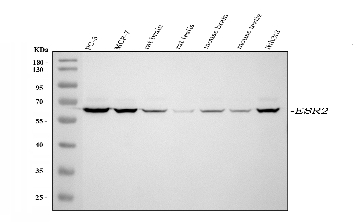
Western blot analysis of ESR2 using anti-ESR2 antibod. The sample well of each lane was loaded with 30 ug of sample under reducing conditions. Lane 1: PC-3 whole cell lysates, Lane 2: MCF-7 whole cell lysates, Lane 3: rat brain tissue lysates, Lane 4: rat testis tissue lysates, Lane 5: mouse brain tissue lysates, Lane 6: mouse testis tissue lysates, Lane 7: NIH/3T3 whole cell lysates. After electrophoresis, proteins were transferred to a membrane. Then the membrane was incubated with rabbit anti-ESR2 antigen affinity purified polyclonal antibody at a dilution of 1:1000 and probed with a goat anti-rabbit IgG-HRP secondary antibody. The signal is developed using ECL Plus Western Blotting Substrate.| Product Name | Rabbit polyclonal antibody to ESR2 |
|---|---|
| Antibody Type | Primary Antibodies |
| Immunogen | E. coli-derived human ESR2 recombinant protein (Position: F289-R501). Human ESR2 shares 93.4% and 93.9% amino acid (aa) sequence identity with mouse and rat ESR2, respectively. |
| Clonality | Polyclonal |
|---|---|
| Isotype | IgG |
| Host Species | Rabbit |
| Tested Applications | WB |
| WB:1:500-1:2000 |
|
| Species Reactivity | HumanMouseRat |
| Concentration | 0.5mg/ml |
| Purification | Affinity purified |
| Gene Symbol | ESR2 |
|---|---|
| Gene Synonyms | Erb ESRB ODG8 ESTRB NR3A2 ER-BETA ESR-BETA |
| Gene Full Name | estrogen receptor 2 |
| Gene Summary | This gene encodes a member of the family of estrogen receptors and superfamily of nuclear receptor transcription factors. The gene product contains an N-terminal DNA binding domain and C-terminal ligand binding domain and is localized to the nucleus, cytoplasm, and mitochondria. Upon binding to 17beta-estradiol or related ligands, the encoded protein forms homo- or hetero-dimers that interact with specific DNA sequences to activate transcription. Some isoforms dominantly inhibit the activity of other estrogen receptor family members. Several alternatively spliced transcript variants of this gene have been described, but the full-length nature of some of these variants has not been fully characterized. [provided by RefSeq, Jul 2008] |
| Alternative Names | ER BETA Erb ESR BETA ESR2 ESRB ESTRB estrogen receptor 2 (ER beta) Estrogen receptor beta Estrogen Receptor β NR3A2 |
| Molecular Weight(MW) | 59 kDa |
WB
Western blot analysis of ESR2 using anti-ESR2 antibod. The sample well of each lane was loaded with 30 ug of sample under reducing conditions. Lane 1: PC-3 whole cell lysates, Lane 2: MCF-7 whole cell lysates, Lane 3: rat brain tissue lysates, Lane 4: rat testis tissue lysates, Lane 5: mouse brain tissue lysates, Lane 6: mouse testis tissue lysates, Lane 7: NIH/3T3 whole cell lysates. After electrophoresis, proteins were transferred to a membrane. Then the membrane was incubated with rabbit anti-ESR2 antigen affinity purified polyclonal antibody at a dilution of 1:1000 and probed with a goat anti-rabbit IgG-HRP secondary antibody. The signal is developed using ECL Plus Western Blotting Substrate.| Application Notes | WB:1:500-1:2000 |
|---|
| Form | Liquid |
|---|---|
| Storage Instructions | 12 months from date of receipt, -20℃ as supplied. 6 months 2 to 8℃ after reconstitution. Avoid repeated freezing and thawing. |
| Storage Buffer | 500ug/ml antibody with PBS, 0.02% NaN3, 1 mg/ml BSA and 50% glycerol. |


