WB
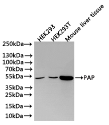
Positive WB detected in: HEK293 whole cell, lysate(30µg), HEK293T whole cell lysate(30µg), Mouse liver tissue lysate(30µg), All lanes: PAP antibody at 1:1000, Secondary, Goat polyclonal to rabbit IgG at 1/40000 dilution, Exposure time:3min.IHC
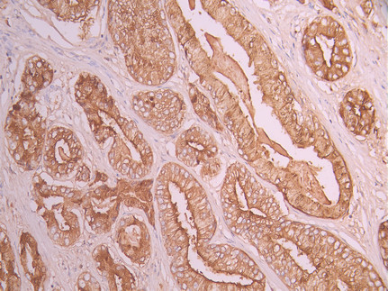
IHC image of ACPP Recombinant Monoclonal Antibody diluted at 1:100 and staining in paraffin-embedded human prostate cancer performed on a Leica BondTM system. After dewaxing and hydration, antigen retrieval was mediated by high pressure in a citrate buffer (pH 6.0). Section was blocked with 10% normal goat serum 30min at RT. Then primary antibody (1% BSA) was incubated at 4°C overnight. The primary is detected by a Goat anti-rabbit polymer IgG labeled by HRP and visualized using 0.05% DAB.| Product Name | ACPP Recombinant Monoclonal Antibody |
|---|---|
| Antibody Type | Primary Antibodies |
| Immunogen | Recombinant Antibody |
| Clonality | Monoclonal |
|---|---|
| Isotype | IgG |
| Host Species | Rabbit |
| Tested Applications | IHCWB |
| WB:1:500-1:5000 IHC:1:50-1:200 |
|
| Species Reactivity | HumanMouse |
| Concentration | 1mg/ml |
| Purification | Affinity purified |
| Gene Symbol | ACP3 |
|---|---|
| Gene Synonyms | ACPP 5'-NT ACP-3 TM-PAP |
| Gene Full Name | acid phosphatase 3 |
| Gene Summary | This gene encodes an enzyme that catalyzes the conversion of orthophosphoric monoester to alcohol and orthophosphate. It is synthesized under androgen regulation and is secreted by the epithelial cells of the prostate gland. An alternatively spliced transcript variant encoding a longer isoform has been found for this gene. This isoform contains a transmembrane domain and is localized in the plasma membrane-endosomal-lysosomal pathway. [provided by RefSeq, Sep 2008] |
| Alternative Names | Prostatic acid phosphatase (PAP) (EC 3.1.3.2) (5'-nucleotidase) (5'-NT) (EC 3.1.3.5) (Ecto-5'-nucleotidase) (Thiamine monophosphatase) (TMPase) [Cleaved into: PAPf39] ACPP |
| Molecular Weight(MW) | 45 kDa |
| Cellular Localization | [Isoform 1]: Secreted.; [Isoform 2]: Cell membrane; Single-pass type I membrane protein. Lysosome membrane; Single-pass type I membrane protein. Nucleus. Cytoplasm, cytosol. |
WB
Positive WB detected in: HEK293 whole cell, lysate(30µg), HEK293T whole cell lysate(30µg), Mouse liver tissue lysate(30µg), All lanes: PAP antibody at 1:1000, Secondary, Goat polyclonal to rabbit IgG at 1/40000 dilution, Exposure time:3min.
IHC
IHC image of ACPP Recombinant Monoclonal Antibody diluted at 1:100 and staining in paraffin-embedded human prostate cancer performed on a Leica BondTM system. After dewaxing and hydration, antigen retrieval was mediated by high pressure in a citrate buffer (pH 6.0). Section was blocked with 10% normal goat serum 30min at RT. Then primary antibody (1% BSA) was incubated at 4°C overnight. The primary is detected by a Goat anti-rabbit polymer IgG labeled by HRP and visualized using 0.05% DAB.| Application Notes | WB:1:500-1:5000 IHC:1:50-1:200 |
|---|
| Form | Liquid |
|---|---|
| Storage Instructions | Upon receipt, store at -20°C or -80°C. Avoid repeated freeze. |
| Storage Buffer | 10mM phosphate buffered saline , pH 7.4, 150mM sodium chloride, 0.05% BSA, 0.02% sodium azide and 50% glycerol. |


