WB
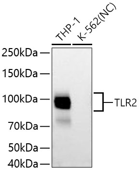
Western blot analysis of various lysates using TLR2 Rabbit pAb at 1:5000 dilution incubated overnight at 4℃. Secondary antibody: HRP-conjugated Goat anti-Rabbit IgG (H+L) at 1:10000 dilution. Lysates/proteins: 25 μg per lane. Blocking buffer: 3% nonfat dry milk in TBST. Detection: ECL Basic Kit. Negative control (NC): K-562. Exposure time: 45 s.IHC
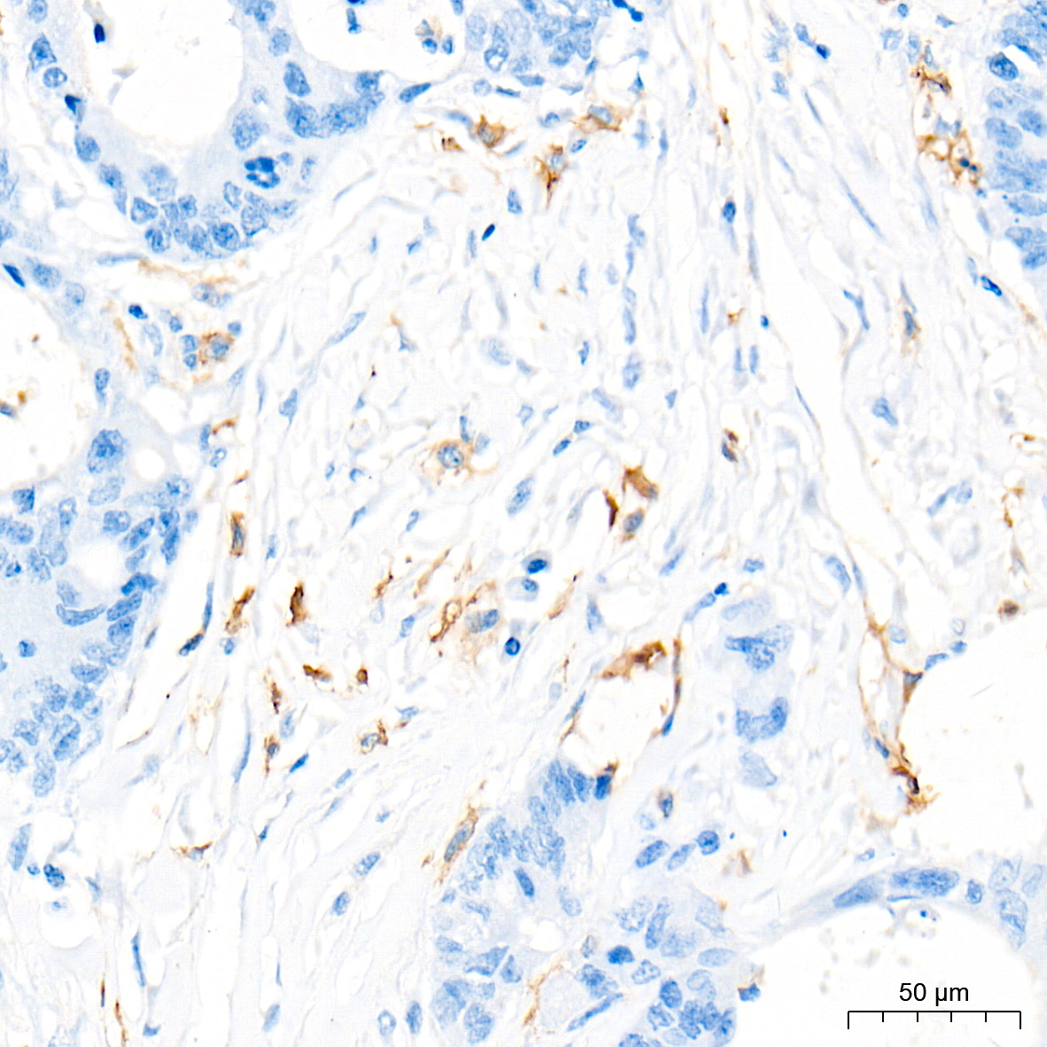
Immunohistochemistry analysis of paraffin-embedded Human colon carcinoma tissue using TLR2 Rabbit pAb at a dilution of 1:5000 (40x lens). High pressure antigen retrieval performed with 0.01M Tris-EDTA Buffer (pH 9.0) prior to IHC staining.IHC
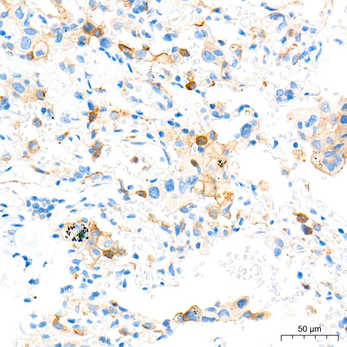
Immunohistochemistry analysis of paraffin-embedded Human lung cancer tissue using TLR2 Rabbit pAb at a dilution of 1:5000 (40x lens). High pressure antigen retrieval performed with 0.01M Tris-EDTA Buffer (pH 9.0) prior to IHC staining.IHC
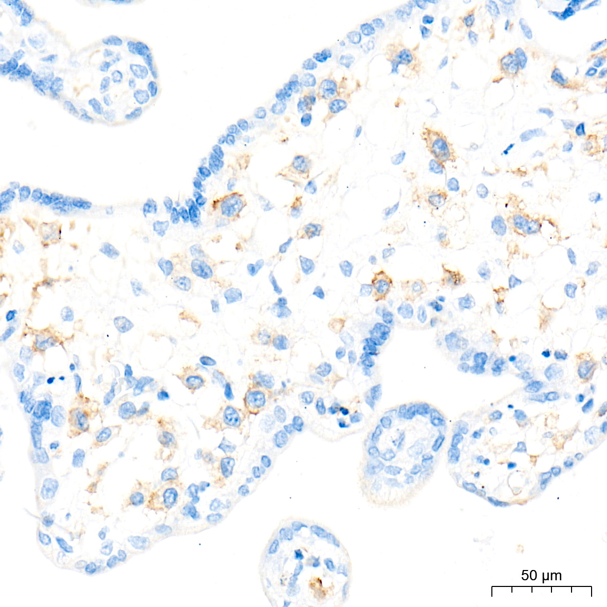
Immunohistochemistry analysis of paraffin-embedded Human placenta tissue using TLR2 Rabbit pAb at a dilution of 1:5000 (40x lens). High pressure antigen retrieval performed with 0.01M Tris-EDTA Buffer (pH 9.0) prior to IHC staining.IHC
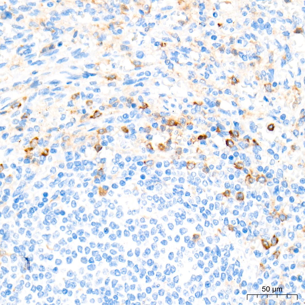
Immunohistochemistry analysis of paraffin-embedded Human spleen tissue using TLR2 Rabbit pAb at a dilution of 1:5000 (40x lens). High pressure antigen retrieval performed with 0.01M Tris-EDTA Buffer (pH 9.0) prior to IHC staining.| Product Name | TLR2 Rabbit pAb |
|---|---|
| Antibody Type | Primary Antibodies |
| Immunogen | This information is considered to be commercially sensitive. |
| Clonality | Polyclonal |
|---|---|
| Isotype | IgG |
| Host Species | Rabbit |
| Tested Applications | IHCWB |
| WB:1:5000-1:10000 IHC:1:1500-1:6000 |
|
| Species Reactivity | HumanMouse |
| Concentration | 1mg/ml |
| Purification | Affinity purified |
| Gene Symbol | TLR2 |
|---|---|
| Gene Synonyms | TIL4 CD282 |
| Gene Full Name | toll like receptor 2 |
| Gene Summary | The protein encoded by this gene is a member of the Toll-like receptor (TLR) family which plays a fundamental role in pathogen recognition and activation of innate immunity. TLRs are highly conserved from Drosophila to humans and share structural and functional similarities. This protein is a cell-surface protein that can form heterodimers with other TLR family members to recognize conserved molecules derived from microorganisms known as pathogen-associated molecular patterns (PAMPs). Activation of TLRs by PAMPs leads to an up-regulation of signaling pathways to modulate the host's inflammatory response. This gene is also thought to promote apoptosis in response to bacterial lipoproteins. This gene has been implicated in the pathogenesis of several autoimmune diseases. Alternative splicing results in multiple transcript variants. [provided by RefSeq, Jan 2016] |
| Alternative Names | TIL4 CD282 TLR2 |
| Molecular Weight(MW) | 90 kDa(Observed band size: 90-105 kDa) |
| Cellular Localization | Membrane, Single-pass type I membrane protein. |
WB
Western blot analysis of various lysates using TLR2 Rabbit pAb at 1:5000 dilution incubated overnight at 4℃. Secondary antibody: HRP-conjugated Goat anti-Rabbit IgG (H+L) at 1:10000 dilution. Lysates/proteins: 25 μg per lane. Blocking buffer: 3% nonfat dry milk in TBST. Detection: ECL Basic Kit. Negative control (NC): K-562. Exposure time: 45 s.
IHC
Immunohistochemistry analysis of paraffin-embedded Human colon carcinoma tissue using TLR2 Rabbit pAb at a dilution of 1:5000 (40x lens). High pressure antigen retrieval performed with 0.01M Tris-EDTA Buffer (pH 9.0) prior to IHC staining.
IHC
Immunohistochemistry analysis of paraffin-embedded Human lung cancer tissue using TLR2 Rabbit pAb at a dilution of 1:5000 (40x lens). High pressure antigen retrieval performed with 0.01M Tris-EDTA Buffer (pH 9.0) prior to IHC staining.
IHC
Immunohistochemistry analysis of paraffin-embedded Human placenta tissue using TLR2 Rabbit pAb at a dilution of 1:5000 (40x lens). High pressure antigen retrieval performed with 0.01M Tris-EDTA Buffer (pH 9.0) prior to IHC staining.
IHC
Immunohistochemistry analysis of paraffin-embedded Human spleen tissue using TLR2 Rabbit pAb at a dilution of 1:5000 (40x lens). High pressure antigen retrieval performed with 0.01M Tris-EDTA Buffer (pH 9.0) prior to IHC staining.| Application Notes | WB:1:5000-1:10000 IHC:1:1500-1:6000 |
|---|
| Form | Liquid |
|---|---|
| Storage Instructions | Store at -20℃. Avoid freeze / thaw cycles. |
| Storage Buffer | Buffer: PBS with 0.05% proclin300, 0.05% BSA, 50% glycerol, pH7.3. |


