WB
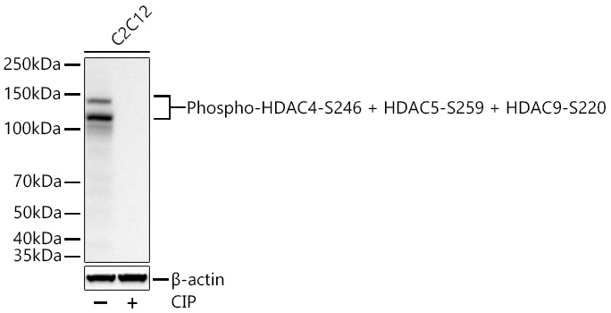
Western blot analysis of lysates from C2C12 cells using Phospho-HDAC4-S246 + HDAC5-S259 + HDAC9-S220 Rabbit mAb at 1:1000 dilution incubated at room temperature for 1.5 hours. C2C12 treated with CIP (1 U/ul) at 37℃ for 1 hour. Secondary antibody: HRP-conjugated Goat anti-Rabbit IgG (H+L) at 1:10000 dilution. Lysates/proteins: 30 μg per lane. Blocking buffer: 3% nonfat dry milk in TBST. Detection: ECL Basic Kit. Exposure time: 30 s.WB
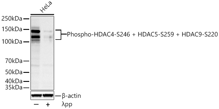
Western blot analysis of lysates from HeLa cells using Phospho-HDAC4-S246 + HDAC5-S259 + HDAC9-S220 Rabbit mAb at 1:1000 dilution incubated at room temperature for 1.5 hours. HeLa treated with λpp (2 U/ul) at 37℃ for 1 hour. Secondary antibody: HRP-conjugated Goat anti-Rabbit IgG (H+L) at 1:10000 dilution. Lysates/proteins: 30 μg per lane. Blocking buffer: 3% nonfat dry milk in TBST. Detection: ECL Basic Kit. Exposure time: 60 s.IHC
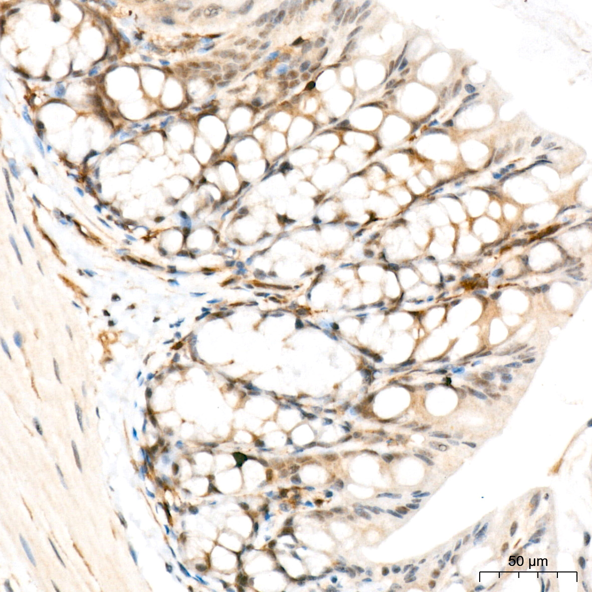
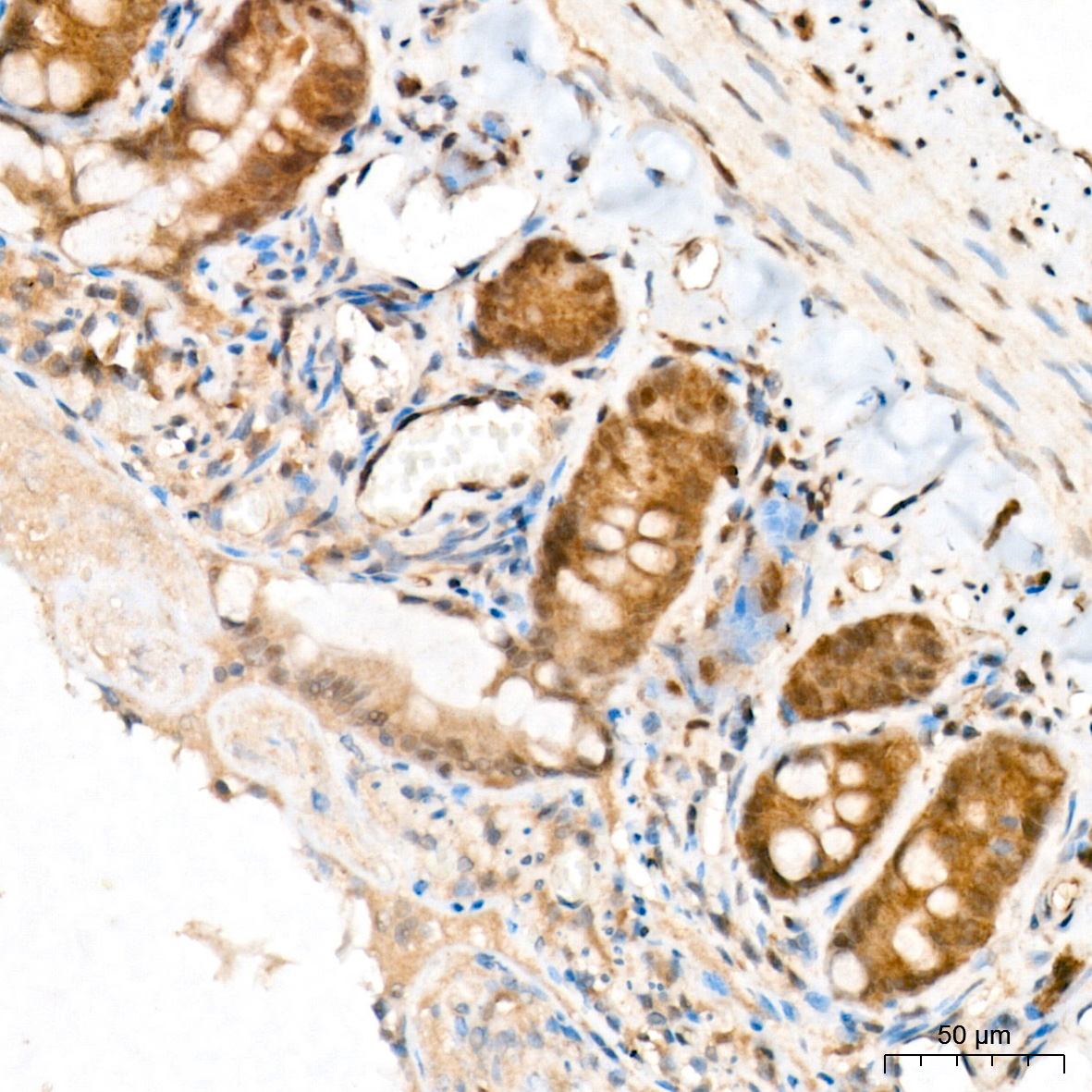
Immunohistochemistry analysis of paraffin-embedded Mouse colon tissue using Phospho-HDAC4-S246 + HDAC5-S259 + HDAC9-S220 Rabbit mAb at a dilution of 1:200 (40x lens). High pressure antigen retrieval performed with 0.01M Citrate Buffer (pH 6.0) prior to IHC staining.IHC
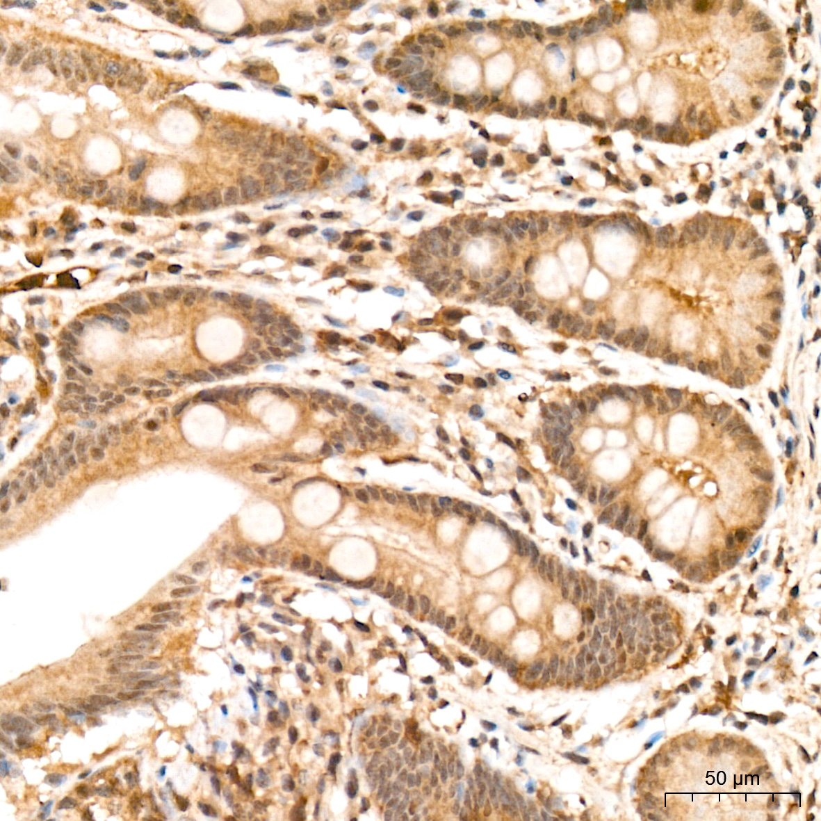
Immunohistochemistry analysis of paraffin-embedded Human colon tissue using Phospho-HDAC4-S246 + HDAC5-S259 + HDAC9-S220 Rabbit mAb at a dilution of 1:200 (40x lens). High pressure antigen retrieval performed with 0.01M Citrate Buffer (pH 6.0) prior to IHC staining.IHC
Immunohistochemistry analysis of paraffin-embedded Rat colon tissue using Phospho-HDAC4-S246 + HDAC5-S259 + HDAC9-S220 Rabbit mAb at a dilution of 1:200 (40x lens). High pressure antigen retrieval performed with 0.01M Citrate Buffer (pH 6.0) prior to IHC staining.| Product Name | Phospho-HDAC4-S246 + HDAC5-S259 + HDAC9-S220 Rabbit mAb |
|---|---|
| Antibody Type | Primary Antibodies |
| Immunogen | Synthetic peptide. This information is considered to be commercially sensitive. |
| Modification | p-S246+S259+S220 |
| Clonality | Monoclonal |
|---|---|
| Isotype | IgG |
| Host Species | Rabbit |
| Tested Applications | IHCWB |
| WB:1:500-1:1000 IHC:1:100-1:400 |
|
| Species Reactivity | HumanMouseRat |
| Concentration | 1mg/ml |
| Purification | Affinity purified |
| Gene Symbol | HDAC4 |
|---|---|
| Gene Synonyms | HD4 AHO3 BDMR HDACA HA6116 HDAC-4 HDAC-A NEDCHF NEDCHID |
| Gene Full Name | histone deacetylase 4 |
| Gene Summary | Histones play a critical role in transcriptional regulation, cell cycle progression, and developmental events. Histone acetylation/deacetylation alters chromosome structure and affects transcription factor access to DNA. The protein encoded by this gene belongs to class II of the histone deacetylase/acuc/apha family. It possesses histone deacetylase activity and represses transcription when tethered to a promoter. This protein does not bind DNA directly, but through transcription factors MEF2C and MEF2D. It seems to interact in a multiprotein complex with RbAp48 and HDAC3. [provided by RefSeq, Jul 2008] |
| Alternative Names | HDAC4/HDAC5/HDAC9 Phospho-HDAC4-S246 + HDAC5-S259 + HDAC9-S220 |
| Molecular Weight(MW) | 57-65 kDa/97-122 kDa(Observed band size: 124 kDa/140 kDa) |
| Cellular Localization | cytoplasm, cytosol, histone deacetylase complex, nuclear speck, nucleoplasm, nucleus. |
WB
Western blot analysis of lysates from C2C12 cells using Phospho-HDAC4-S246 + HDAC5-S259 + HDAC9-S220 Rabbit mAb at 1:1000 dilution incubated at room temperature for 1.5 hours. C2C12 treated with CIP (1 U/ul) at 37℃ for 1 hour. Secondary antibody: HRP-conjugated Goat anti-Rabbit IgG (H+L) at 1:10000 dilution. Lysates/proteins: 30 μg per lane. Blocking buffer: 3% nonfat dry milk in TBST. Detection: ECL Basic Kit. Exposure time: 30 s.
WB
Western blot analysis of lysates from HeLa cells using Phospho-HDAC4-S246 + HDAC5-S259 + HDAC9-S220 Rabbit mAb at 1:1000 dilution incubated at room temperature for 1.5 hours. HeLa treated with λpp (2 U/ul) at 37℃ for 1 hour. Secondary antibody: HRP-conjugated Goat anti-Rabbit IgG (H+L) at 1:10000 dilution. Lysates/proteins: 30 μg per lane. Blocking buffer: 3% nonfat dry milk in TBST. Detection: ECL Basic Kit. Exposure time: 60 s.
IHC
Immunohistochemistry analysis of paraffin-embedded Mouse colon tissue using Phospho-HDAC4-S246 + HDAC5-S259 + HDAC9-S220 Rabbit mAb at a dilution of 1:200 (40x lens). High pressure antigen retrieval performed with 0.01M Citrate Buffer (pH 6.0) prior to IHC staining.
IHC
Immunohistochemistry analysis of paraffin-embedded Human colon tissue using Phospho-HDAC4-S246 + HDAC5-S259 + HDAC9-S220 Rabbit mAb at a dilution of 1:200 (40x lens). High pressure antigen retrieval performed with 0.01M Citrate Buffer (pH 6.0) prior to IHC staining.
IHC
Immunohistochemistry analysis of paraffin-embedded Rat colon tissue using Phospho-HDAC4-S246 + HDAC5-S259 + HDAC9-S220 Rabbit mAb at a dilution of 1:200 (40x lens). High pressure antigen retrieval performed with 0.01M Citrate Buffer (pH 6.0) prior to IHC staining.| Application Notes | WB:1:500-1:1000 IHC:1:100-1:400 |
|---|
| Form | Liquid |
|---|---|
| Storage Instructions | Store at -20℃. Avoid freeze / thaw cycles. |
| Storage Buffer | Buffer: PBS with 0.05% proclin300, 0.05% BSA, 50% glycerol, pH7.3. |


