

WB
Positive WB detected in:A-431 whole cell lysate(30µg), PC-3 whole cell lysate(30µg), Hela whole cell lysate(30µg), jurkat whole cell lysate(30µg), HepG2 whole cell lysate(30µg), A549 whole cell lysate(30µg), U251 whole cell lysate(30µg).All lanes: Lipoma preferred partner antibody at 1:1000.Secondary,Goat polyclonal to rabbit IgG at 1/40000 dilution.Exposure time:60s.IHC
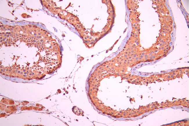
IHC image of PGK1 Recombinant Monoclonal Antibody diluted at 1:100 and staining in paraffin-embedded human testis tissue performed on a Leica BondTM system. After dewaxing and hydration, antigen retrieval was mediated by high pressure in a citrate buffer (pH 6.0). Section was blocked with 10% normal goat serum 30min at RT. Then primary antibody (1% BSA) was incubated at 4°C overnight. The primary is detected by a Goat anti-rabbit polymer IgG labeled by HRP and visualized using 0.05% DAB.IHC
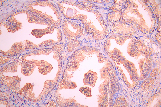
IHC image of PGK1 Recombinant Monoclonal Antibody diluted at 1:100 and staining in paraffin-embedded human prostate cancer performed on a Leica BondTM system. After dewaxing and hydration, antigen retrieval was mediated by high pressure in a citrate buffer (pH 6.0). Section was blocked with 10% normal goat serum 30min at RT. Then primary antibody (1% BSA) was incubated at 4°C overnight. The primary is detected by a Goat anti-rabbit polymer IgG labeled by HRP and visualized using 0.05% DAB.FC
Overlay Peak curve showing 786-O cells stained with PGK1 Recombinant Monoclonal Antibody (red line) at 1:100. The cells were fixed in 4% formaldehyde and permeated by 0.2% TritonX-100 for 10min. Then 10% normal goat serum to block non-specific protein-protein interactions followed by the antibody (1ug/1*106cells) for 45min at 4℃. The secondary antibody used was FITC-conjugated goat anti-rabbit IgG (H+L) at 1/200 dilution for 35min at 4℃.Control antibody (green line) was Rabbit IgG (1ug/1*106cells) used under the same conditions. Acquisition of >10,000 events was performed.| Product Name | PGK1 Recombinant Monoclonal Antibody |
|---|---|
| Antibody Type | Primary Antibodies |
| Immunogen | A synthesized peptide derived from Human PGK1 protein |
| Clonality | Monoclonal |
|---|---|
| Isotype | IgG |
| Host Species | Rabbit |
| Tested Applications | FCIHCWB |
| WB:1:500-1:5000 IHC:1:50-1:200 FC:1:20-1:200 |
|
| Species Reactivity | Human |
| Concentration | 1mg/ml |
| Purification | Affinity purified |
| Gene Symbol | PGK1 |
|---|---|
| Gene Synonyms | PGKA MIG10 HEL-S-68p |
| Gene Full Name | phosphoglycerate kinase 1 |
| Gene Summary | The protein encoded by this gene is a glycolytic enzyme that catalyzes the conversion of 1,3-diphosphoglycerate to 3-phosphoglycerate. The encoded protein may also act as a cofactor for polymerase alpha. Additionally, this protein is secreted by tumor cells where it participates in angiogenesis by functioning to reduce disulfide bonds in the serine protease, plasmin, which consequently leads to the release of the tumor blood vessel inhibitor angiostatin. The encoded protein has been identified as a moonlighting protein based on its ability to perform mechanistically distinct functions. Deficiency of the enzyme is associated with a wide range of clinical phenotypes hemolytic anemia and neurological impairment. Pseudogenes of this gene have been defined on chromosomes 19, 21 and the X chromosome. [provided by RefSeq, Jan 2014] |
| Alternative Names | Phosphoglycerate kinase 1 (EC 2.7.2.3) (Cell migration-inducing gene 10 protein) (Primer recognition protein 2) (PRP 2) PGK1 PGKA |
| Molecular Weight(MW) | 45 kDa(Observed band size: 44 kDa) |
| Cellular Localization | Cytoplasm |
WB
Positive WB detected in:A-431 whole cell lysate(30µg), PC-3 whole cell lysate(30µg), Hela whole cell lysate(30µg), jurkat whole cell lysate(30µg), HepG2 whole cell lysate(30µg), A549 whole cell lysate(30µg), U251 whole cell lysate(30µg).All lanes: Lipoma preferred partner antibody at 1:1000.Secondary,Goat polyclonal to rabbit IgG at 1/40000 dilution.Exposure time:60s.
IHC
IHC image of PGK1 Recombinant Monoclonal Antibody diluted at 1:100 and staining in paraffin-embedded human testis tissue performed on a Leica BondTM system. After dewaxing and hydration, antigen retrieval was mediated by high pressure in a citrate buffer (pH 6.0). Section was blocked with 10% normal goat serum 30min at RT. Then primary antibody (1% BSA) was incubated at 4°C overnight. The primary is detected by a Goat anti-rabbit polymer IgG labeled by HRP and visualized using 0.05% DAB.
IHC
IHC image of PGK1 Recombinant Monoclonal Antibody diluted at 1:100 and staining in paraffin-embedded human prostate cancer performed on a Leica BondTM system. After dewaxing and hydration, antigen retrieval was mediated by high pressure in a citrate buffer (pH 6.0). Section was blocked with 10% normal goat serum 30min at RT. Then primary antibody (1% BSA) was incubated at 4°C overnight. The primary is detected by a Goat anti-rabbit polymer IgG labeled by HRP and visualized using 0.05% DAB.
FC
Overlay Peak curve showing 786-O cells stained with PGK1 Recombinant Monoclonal Antibody (red line) at 1:100. The cells were fixed in 4% formaldehyde and permeated by 0.2% TritonX-100 for 10min. Then 10% normal goat serum to block non-specific protein-protein interactions followed by the antibody (1ug/1*106cells) for 45min at 4℃. The secondary antibody used was FITC-conjugated goat anti-rabbit IgG (H+L) at 1/200 dilution for 35min at 4℃.Control antibody (green line) was Rabbit IgG (1ug/1*106cells) used under the same conditions. Acquisition of >10,000 events was performed.| Application Notes | WB:1:500-1:5000 IHC:1:50-1:200 FC:1:20-1:200 |
|---|
| Form | Liquid |
|---|---|
| Storage Instructions | Upon receipt, store at -20°C or -80°C. Avoid repeated freeze. |
| Storage Buffer | 0.03% Proclin 300 Constituents: 50% Glycerol, 0.01M PBS, PH 7.4 |


