WB
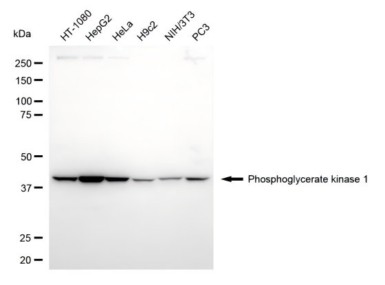
Western blotting analysis using phosphoglycerate kinase 1 antibody. Total cell lysates (30 μg) from various cell lines were loaded and separated by SDS-PAGE. The blot was incubated with phosphoglycerate kinase 1 antibody and HRP-conjugated goat anti-rabbit secondary antibody respectively.ICC/IF
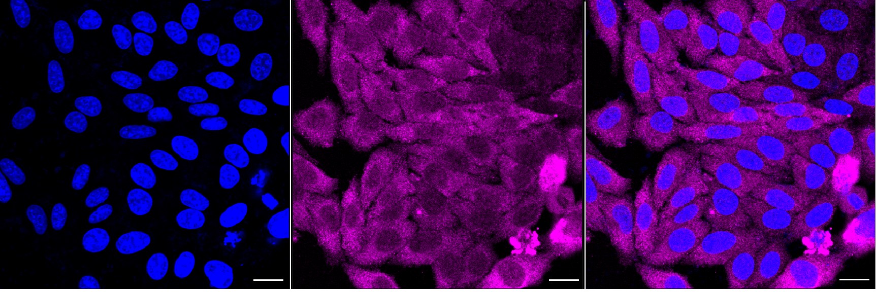
Immunocytochemical staining of HepG2 cells with Phosphoglycerate kinase 1 antibody. Nuclei were stained blue with DAPI; Phosphoglycerate kinase 1 was stained magenta with Alexa Fluor® 647. Images were taken using Leica stellaris 5. Protein abundance based on laser Intensity and smart gain: Medium. Scale bar, 20 μm.FC
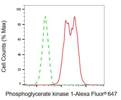
Flow cytometric analysis of phosphoglycerate kinase 1 expression in HAP-1 cells using phosphoglycerate kinase 1 antibody. Green, isotype control; red, phosphoglycerate kinase 1.| Product Name | PGK1 Recombinant Monoclonal Antibody |
|---|---|
| Antibody Type | Primary Antibodies |
| Immunogen | Recombinant Human PGK1 protein |
| Clonality | Monoclonal |
|---|---|
| Isotype | IgG |
| Host Species | Rabbit |
| Tested Applications | FCICC/IFWB |
| WB:1:1000-1:5000 ICC/IF:1:100-1:1000 FC:1:200-1:2000 |
|
| Species Reactivity | HumanMouseRabbit |
| Concentration | 1mg/ml |
| Purification | Affinity purified |
| Gene Symbol | PGK1 |
|---|---|
| Gene Synonyms | PGKA MIG10 HEL-S-68p |
| Gene Full Name | phosphoglycerate kinase 1 |
| Gene Summary | The protein encoded by this gene is a glycolytic enzyme that catalyzes the conversion of 1,3-diphosphoglycerate to 3-phosphoglycerate. The encoded protein may also act as a cofactor for polymerase alpha. Additionally, this protein is secreted by tumor cells where it participates in angiogenesis by functioning to reduce disulfide bonds in the serine protease, plasmin, which consequently leads to the release of the tumor blood vessel inhibitor angiostatin. The encoded protein has been identified as a moonlighting protein based on its ability to perform mechanistically distinct functions. Deficiency of the enzyme is associated with a wide range of clinical phenotypes hemolytic anemia and neurological impairment. Pseudogenes of this gene have been defined on chromosomes 19, 21 and the X chromosome. [provided by RefSeq, Jan 2014] |
| Alternative Names | PGK1 Phosphoglycerate Kinase 1 Cell Migration-Inducing Gene 10 Protein Primer Recognition Protein EC 2.7.2.3 PRP PGKA Epididymis Secretory Sperm Binding Protein Li 68p HEL-S-68p MIG10 |
| Cellular Localization | Cytoplasm |
WB
Western blotting analysis using phosphoglycerate kinase 1 antibody. Total cell lysates (30 μg) from various cell lines were loaded and separated by SDS-PAGE. The blot was incubated with phosphoglycerate kinase 1 antibody and HRP-conjugated goat anti-rabbit secondary antibody respectively.
ICC/IF
Immunocytochemical staining of HepG2 cells with Phosphoglycerate kinase 1 antibody. Nuclei were stained blue with DAPI; Phosphoglycerate kinase 1 was stained magenta with Alexa Fluor® 647. Images were taken using Leica stellaris 5. Protein abundance based on laser Intensity and smart gain: Medium. Scale bar, 20 μm.
FC
Flow cytometric analysis of phosphoglycerate kinase 1 expression in HAP-1 cells using phosphoglycerate kinase 1 antibody. Green, isotype control; red, phosphoglycerate kinase 1.| Application Notes | WB:1:1000-1:5000 ICC/IF:1:100-1:1000 FC:1:200-1:2000 |
|---|
| Form | Liquid |
|---|---|
| Storage Instructions | Upon receipt, store at -20°C or -80°C. Avoid repeated freeze. |
| Storage Buffer | PBS (pH 7.4) containing 50% glycerol, and 0.02% sodium azide. |


