WB
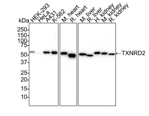
Western blot analysis of TXNRD2 on different lysates with Rabbit anti-TXNRD2 antibody at 1/1,000 dilution. Lane 1: HEK-293 cell lysate (20 µg/Lane), Lane 2: HeLa cell lysate (20 µg/Lane), Lane 3: A431 cell lysate (20 µg/Lane), Lane 4: K-562 cell lysate (20 µg/Lane), Lane 5: Mouse heart tissue lysate (40 µg/Lane), Lane 6: Rat heart tissue lysate (40 µg/Lane), Lane 7: Mouse liver tissue lysate (40 µg/Lane), Lane 8: Rat liver tissue lysate (40 µg/Lane), Lane 9: Human kidney tissue lysate (40 µg/Lane), Lane 10: Mouse kidney tissue lysate (40 µg/Lane), Lane 11: Rat kidney tissue lysate (40 µg/Lane), Exposure time: 3 minutes; 4-20% SDS-PAGE gel. Proteins were transferred to a PVDF membrane and blocked with 5% NFDM/TBST for 1 hour at room temperature. The primary antibody at 1/1,000 dilution a was used in 5% NFDM/TBST at 4℃ overnight. Goat Anti-Rabbit IgG - HRP Secondary Antibody at 1/50,000 dilution was used for 1 hour at room temperature.IHC
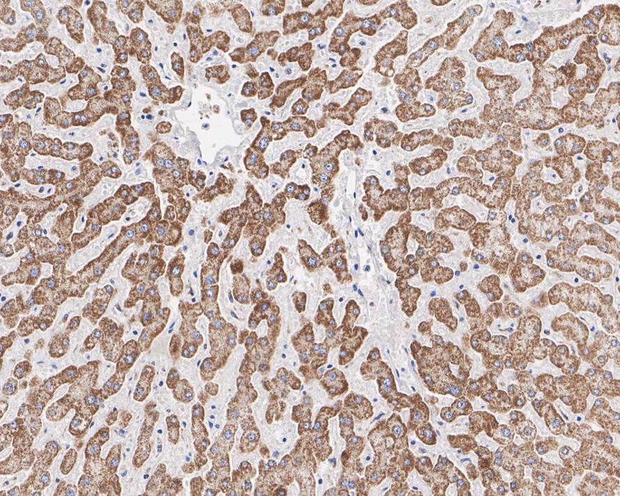
Immunohistochemical analysis of paraffin-embedded human liver tissue with Rabbit anti-TXNRD2 antibody at 1/1,000 dilution. The section was pre-treated using heat mediated antigen retrieval with Tris-EDTA buffer (pH 9.0) for 20 minutes. The tissues were blocked in 1% BSA for 20 minutes at room temperature, washed with ddH2O and PBS, and then probed with the primary antibody at 1/1,000 dilution for 1 hour at room temperature. The detection was performed using an HRP conjugated compact polymer system. DAB was used as the chromogen. Tissues were counterstained with hematoxylin and mounted with DPX.IHC
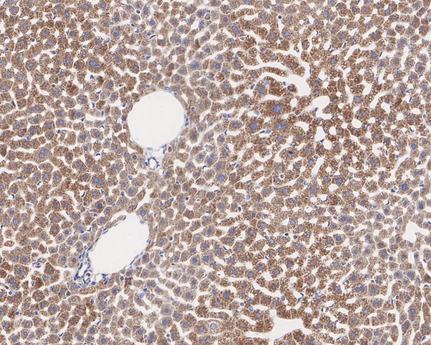
Immunohistochemical analysis of paraffin-embedded mouse liver tissue with Rabbit anti-TXNRD2 antibody at 1/200 dilution. The section was pre-treated using heat mediated antigen retrieval with Tris-EDTA buffer (pH 9.0) for 20 minutes. The tissues were blocked in 1% BSA for 20 minutes at room temperature, washed with ddH2O and PBS, and then probed with the primary antibody (HA721806) at 1/200 dilution for 1 hour at room temperature. The detection was performed using an HRP conjugated compact polymer system. DAB was used as the chromogen. Tissues were counterstained with hematoxylin and mounted with DPX.IHC
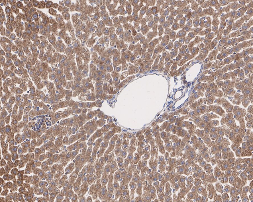
Immunohistochemical analysis of paraffin-embedded rat liver tissue with Rabbit anti-TXNRD2 antibody at 1/200 dilution. The section was pre-treated using heat mediated antigen retrieval with Tris-EDTA buffer (pH 9.0) for 20 minutes. The tissues were blocked in 1% BSA for 20 minutes at room temperature, washed with ddH2O and PBS, and then probed with the primary antibody at 1/200 dilution for 1 hour at room temperature. The detection was performed using an HRP conjugated compact polymer system. DAB was used as the chromogen. Tissues were counterstained with hematoxylin and mounted with DPX.IF-P
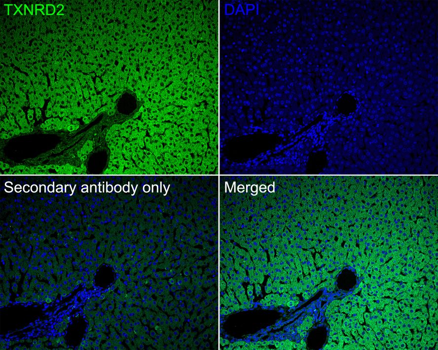
Immunofluorescence analysis of paraffin-embedded rat liver tissue labeling TXNRD2 with Rabbit anti-TXNRD2 antibody at 1/50 dilution. The section was pre-treated using heat mediated antigen retrieval with Tris-EDTA buffer (pH 9.0) for 20 minutes. The tissues were blocked in 10% negative goat serum for 1 hour at room temperature, washed with PBS, and then probed with the primary antibody (green) at 1/50 dilution overnight at 4 ℃, washed with PBS. Goat Anti-Rabbit IgG H&L (488) was used as the secondary antibody at 1/1,000 dilution. Nuclei were counterstained with DAPI (blue).| Product Name | TXNRD2 Recombinant Rabbit Monoclonal Antibody |
|---|---|
| Antibody Type | Primary Antibodies |
| Immunogen | Recombinant protein within human TXNRD2 aa 1-524 / 524. |
| Clonality | Monoclonal |
|---|---|
| Isotype | IgG |
| Host Species | Rabbit |
| Tested Applications | IF-PIHCWB |
| WB:1:1000 IHC:1:200-1:1000 IF-P:1:50 |
|
| Species Reactivity | HumanMouseRat |
| Concentration | 1mg/ml |
| Purification | Protein A |
| Gene Symbol | TXNRD2 |
|---|---|
| Gene Synonyms | TR TR3 SELZ GCCD5 TRXR2 TXNR2 TR-BETA |
| Gene Full Name | thioredoxin reductase 2 |
| Gene Summary | The protein encoded by this gene belongs to the pyridine nucleotide-disulfide oxidoreductase family, and is a member of the thioredoxin (Trx) system. Three thioredoxin reductase (TrxR) isozymes are found in mammals. TrxRs are selenocysteine-containing flavoenzymes, which reduce thioredoxins, as well as other substrates, and play a key role in redox homoeostasis. This gene encodes a mitochondrial form important for scavenging reactive oxygen species in mitochondria. It functions as a homodimer containing FAD, and selenocysteine (Sec) at the active site. Sec is encoded by UGA codon that normally signals translation termination. The 3' UTRs of selenoprotein mRNAs contain a conserved stem-loop structure, the Sec insertion sequence (SECIS) element, which is necessary for the recognition of UGA as a Sec codon rather than as a stop signal. Alternatively spliced transcript variants encoding different isoforms, including a few localized in the cytosol and some lacking the C-terminal Sec residue, have been found for this gene. [provided by RefSeq, Jun 2017] |
| Alternative Names | mitochondrial antibody Selenoprotein Z antibody SelZ antibody Thioredoxin reductase 2 antibody Thioredoxin reductase 2 mitochondrial antibody Thioredoxin reductase 3 antibody Thioredoxin reductase beta antibody Thioredoxin reductase TR3 antibody TR 3 antibody TR antibody mitochondrial antibody Selenoprotein Z antibody SelZ antibody Thioredoxin reductase 2 antibody Thioredoxin reductase 2 mitochondrial antibody Thioredoxin reductase 3 antibody Thioredoxin reductase beta antibody Thioredoxin reductase TR3 antibody TR 3 antibody TR antibody TR beta antibody TR-beta antibody TR3 antibody TRXR 2 antibody TRXR2 antibody TRXR2_HUMAN antibody TXNRD 2 antibody Txnrd2 antibody |
| Molecular Weight(MW) | 57 kDa |
| Cellular Localization | Mitochondrion. |
WB
Western blot analysis of TXNRD2 on different lysates with Rabbit anti-TXNRD2 antibody at 1/1,000 dilution. Lane 1: HEK-293 cell lysate (20 µg/Lane), Lane 2: HeLa cell lysate (20 µg/Lane), Lane 3: A431 cell lysate (20 µg/Lane), Lane 4: K-562 cell lysate (20 µg/Lane), Lane 5: Mouse heart tissue lysate (40 µg/Lane), Lane 6: Rat heart tissue lysate (40 µg/Lane), Lane 7: Mouse liver tissue lysate (40 µg/Lane), Lane 8: Rat liver tissue lysate (40 µg/Lane), Lane 9: Human kidney tissue lysate (40 µg/Lane), Lane 10: Mouse kidney tissue lysate (40 µg/Lane), Lane 11: Rat kidney tissue lysate (40 µg/Lane), Exposure time: 3 minutes; 4-20% SDS-PAGE gel. Proteins were transferred to a PVDF membrane and blocked with 5% NFDM/TBST for 1 hour at room temperature. The primary antibody at 1/1,000 dilution a was used in 5% NFDM/TBST at 4℃ overnight. Goat Anti-Rabbit IgG - HRP Secondary Antibody at 1/50,000 dilution was used for 1 hour at room temperature.
IHC
Immunohistochemical analysis of paraffin-embedded human liver tissue with Rabbit anti-TXNRD2 antibody at 1/1,000 dilution. The section was pre-treated using heat mediated antigen retrieval with Tris-EDTA buffer (pH 9.0) for 20 minutes. The tissues were blocked in 1% BSA for 20 minutes at room temperature, washed with ddH2O and PBS, and then probed with the primary antibody at 1/1,000 dilution for 1 hour at room temperature. The detection was performed using an HRP conjugated compact polymer system. DAB was used as the chromogen. Tissues were counterstained with hematoxylin and mounted with DPX.
IHC
Immunohistochemical analysis of paraffin-embedded mouse liver tissue with Rabbit anti-TXNRD2 antibody at 1/200 dilution. The section was pre-treated using heat mediated antigen retrieval with Tris-EDTA buffer (pH 9.0) for 20 minutes. The tissues were blocked in 1% BSA for 20 minutes at room temperature, washed with ddH2O and PBS, and then probed with the primary antibody (HA721806) at 1/200 dilution for 1 hour at room temperature. The detection was performed using an HRP conjugated compact polymer system. DAB was used as the chromogen. Tissues were counterstained with hematoxylin and mounted with DPX.
IHC
Immunohistochemical analysis of paraffin-embedded rat liver tissue with Rabbit anti-TXNRD2 antibody at 1/200 dilution. The section was pre-treated using heat mediated antigen retrieval with Tris-EDTA buffer (pH 9.0) for 20 minutes. The tissues were blocked in 1% BSA for 20 minutes at room temperature, washed with ddH2O and PBS, and then probed with the primary antibody at 1/200 dilution for 1 hour at room temperature. The detection was performed using an HRP conjugated compact polymer system. DAB was used as the chromogen. Tissues were counterstained with hematoxylin and mounted with DPX.
IF-P
Immunofluorescence analysis of paraffin-embedded rat liver tissue labeling TXNRD2 with Rabbit anti-TXNRD2 antibody at 1/50 dilution. The section was pre-treated using heat mediated antigen retrieval with Tris-EDTA buffer (pH 9.0) for 20 minutes. The tissues were blocked in 10% negative goat serum for 1 hour at room temperature, washed with PBS, and then probed with the primary antibody (green) at 1/50 dilution overnight at 4 ℃, washed with PBS. Goat Anti-Rabbit IgG H&L (488) was used as the secondary antibody at 1/1,000 dilution. Nuclei were counterstained with DAPI (blue).| Application Notes | WB:1:1000 IHC:1:200-1:1000 IF-P:1:50 |
|---|
| Form | Liquid |
|---|---|
| Storage Instructions | Shipped at 4℃. Store at +4℃ short term (1-2 weeks). It is recommended to aliquot into single-use upon delivery. Store at -20℃ long term. |
| Storage Buffer | 1*TBS (pH7.4), 0.05% BSA, 40% Glycerol. Preservative: 0.05% Sodium Azide. |


