WB
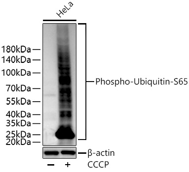
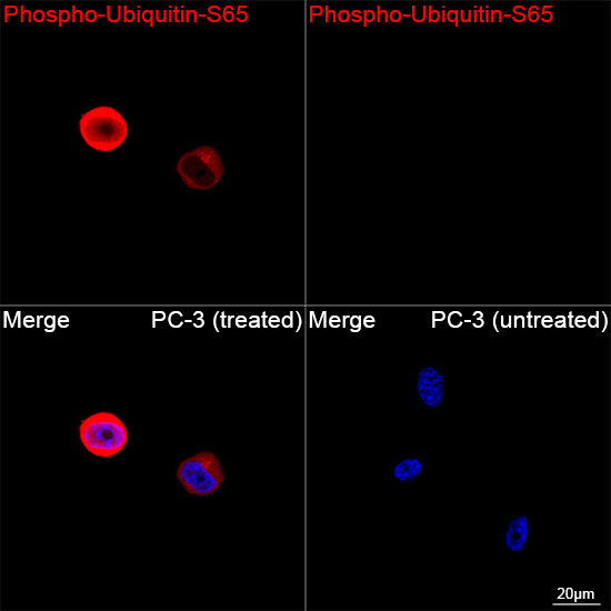
Western blot analysis of lysates from HeLa cells using Phospho-Ubiquitin-S65 Rabbit mAb at 1:5000 dilution incubated overnight at 4℃. HeLa cells were treated with CCCP (30 μM) at 37℃ for 6 hours. Secondary antibody: HRP-conjugated Goat anti-Rabbit IgG (H+L) at 1:10000 dilution. Lysates/proteins: 30 μg per lane. Blocking buffer: 3% nonfat dry milk in TBST. Detection: ECL Basic Kit. Exposure time: 60 s.Confocal imaging of PC-3 cells (treated with CCCP) and PC-3 cells (untreated) using Phospho-Ubiquitin-S65 Rabbit mAb (dilution 1:200) followed by a further incubation with Cy3-conjugated Goat anti-Rabbit IgG (H+L) ( dilution 1:500) (Red). DAPI was used for nuclear staining (Blue). Objective: 100x.
| Product Name | Phospho-Ubiquitin-S65 Rabbit mAb |
|---|---|
| Antibody Type | Primary Antibodies |
| Immunogen | Synthetic peptide. This information is considered to be commercially sensitive. |
| Modification | p-S65 |
| Clonality | Monoclonal |
|---|---|
| Isotype | IgG |
| Host Species | Rabbit |
| Tested Applications | ICC/IFWB |
| WB:1:1000-1:5000 ICC/IF:1:200-1:400 |
|
| Species Reactivity | Human |
| Concentration | 1mg/ml |
| Purification | Affinity purified |
| Gene Symbol | RPS27A |
|---|---|
| Gene Synonyms | S27A eS31 CEP80 UBA80 HEL112 UBCEP1 UBCEP80 |
| Gene Full Name | ribosomal protein S27a |
| Gene Summary | Ubiquitin, a highly conserved protein that has a major role in targeting cellular proteins for degradation by the 26S proteosome, is synthesized as a precursor protein consisting of either polyubiquitin chains or a single ubiquitin fused to an unrelated protein. This gene encodes a fusion protein consisting of ubiquitin at the N terminus and ribosomal protein S27a at the C terminus. When expressed in yeast, the protein is post-translationally processed, generating free ubiquitin monomer and ribosomal protein S27a. Ribosomal protein S27a is a component of the 40S subunit of the ribosome and belongs to the S27AE family of ribosomal proteins. It contains C4-type zinc finger domains and is located in the cytoplasm. Pseudogenes derived from this gene are present in the genome. As with ribosomal protein S27a, ribosomal protein L40 is also synthesized as a fusion protein with ubiquitin; similarly, ribosomal protein S30 is synthesized as a fusion protein with the ubiquitin-like protein fubi. Multiple alternatively spliced transcript variants that encode the same proteins have been identified.[provided by RefSeq, Sep 2008] |
| Alternative Names | UBC S27A eS31 CEP80 UBA80 HEL112 UBCEP1 UBCEP80 L40 CEP52 RPL40 HUBCEP52 HMG20 Phospho-Ubiquitin-Ser65 |
| Molecular Weight(MW) | 18 kDa/15 kDa/26 kDa/77 kDa (Observed band size: 20kDa) |
| Cellular Localization | Cytoplasm, Nucleus, nucleolus, Mitochondrion outer membrane. |
WB
Western blot analysis of lysates from HeLa cells using Phospho-Ubiquitin-S65 Rabbit mAb at 1:5000 dilution incubated overnight at 4℃. HeLa cells were treated with CCCP (30 μM) at 37℃ for 6 hours. Secondary antibody: HRP-conjugated Goat anti-Rabbit IgG (H+L) at 1:10000 dilution. Lysates/proteins: 30 μg per lane. Blocking buffer: 3% nonfat dry milk in TBST. Detection: ECL Basic Kit. Exposure time: 60 s.
Confocal imaging of PC-3 cells (treated with CCCP) and PC-3 cells (untreated) using Phospho-Ubiquitin-S65 Rabbit mAb (dilution 1:200) followed by a further incubation with Cy3-conjugated Goat anti-Rabbit IgG (H+L) ( dilution 1:500) (Red). DAPI was used for nuclear staining (Blue). Objective: 100x.
| Application Notes | WB:1:1000-1:5000 ICC/IF:1:200-1:400 |
|---|
| Form | Liquid |
|---|---|
| Storage Instructions | Store at -20℃. Avoid freeze / thaw cycles. |
| Storage Buffer | Buffer: PBS with 0.09% Sodium azide, 0.05% BSA, 50% glycerol, pH7.3. |


