WB
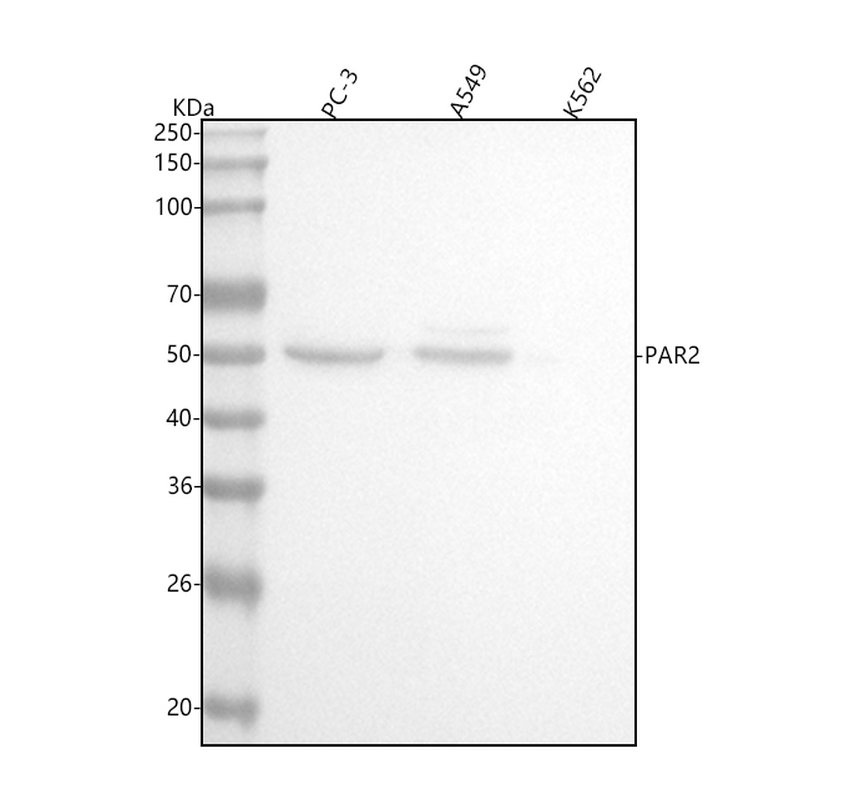
Western blot analysis of anti-PAR2/F2RL1 antibody. The sample well of each lane was loaded with 30 ug of sample under reducing conditions. After electrophoresis, proteins were transferred to a membrane. Then the membrane was incubated with rabbit anti-PAR2/F2RL1 antigen affinity purified monoclonal antibody at a dilution of 1:1000 and probed with a goat anti-rabbit IgG-HRP secondary antibody. The signal is developed using ECL Plus Western Blotting Substrate.IHC
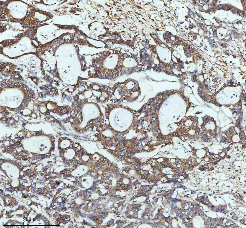
IHC analysis of PAR2/F2RL1 using anti-PAR2/F2RL1 antibody. PAR2/F2RL1 was detected in a paraffin-embedded section of human colon adenocarcinoma tissue. The tissue section was incubated with rabbit anti-PAR2/F2RL1 Antibody at a dilution of 1:200 and developed using HRP Conjugated Rabbit IgG Super Vision Assay Kit with DAB as the chromogen.IHC
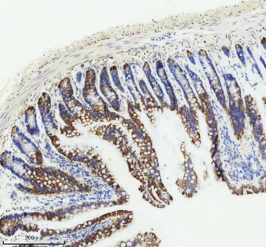
IHC analysis of PAR2/F2RL1 using anti-PAR2/F2RL1 antibody. PAR2/F2RL1 was detected in a paraffin-embedded section of rat small intestine tissue. The tissue section was incubated with rabbit anti-PAR2/F2RL1 Antibody at a dilution of 1:200 and developed using HRP Conjugated Rabbit IgG Super Vision Assay Kit with DAB as the chromogen.| Product Name | Rabbit monoclonal antibody to PAR2/F2RL1 |
|---|---|
| Antibody Type | Primary Antibodies |
| Immunogen | A synthesized peptide derived from human PAR2 |
| Clonality | Monoclonal |
|---|---|
| Isotype | IgG |
| Host Species | Rabbit |
| Tested Applications | IHCWB |
| WB:1:500-1:2000 IHC:1:50-1:200 ICC/IF:1:50-1:200 FC:1:50 |
|
| Species Reactivity | HumanMouseRat |
| Concentration | 0.5mg/ml |
| Purification | Affinity purified |
| Gene Symbol | F2RL1 |
|---|---|
| Gene Synonyms | PAR2 GPR11 |
| Gene Full Name | F2R like trypsin receptor 1 |
| Gene Summary | This gene encodes a member of the G-protein coupled receptor 1 family of proteins. The encoded cell surface receptor is activated through proteolytic cleavage of its extracellular amino terminus, resulting in a new amino terminus that acts as a tethered ligand that binds to an extracellular loop domain. Activation of the receptor has been shown to stimulate vascular smooth muscle relaxation, dilate blood vessels, increase blood flow, and lower blood pressure. This protein is also important in the inflammatory response, as well as innate and adaptive immunity. [provided by RefSeq, Jun 2016] |
| Alternative Names | F2RL1 G protein coupled receptor 11 GPR11 PAR 2 PAR2 Thrombin receptor like 1 |
| Molecular Weight(MW) | 44 kDa(Observed band size: 50 kDa) |
| Cellular Localization | Cell membrane,Membrane. |
WB
Western blot analysis of anti-PAR2/F2RL1 antibody. The sample well of each lane was loaded with 30 ug of sample under reducing conditions. After electrophoresis, proteins were transferred to a membrane. Then the membrane was incubated with rabbit anti-PAR2/F2RL1 antigen affinity purified monoclonal antibody at a dilution of 1:1000 and probed with a goat anti-rabbit IgG-HRP secondary antibody. The signal is developed using ECL Plus Western Blotting Substrate.
IHC
IHC analysis of PAR2/F2RL1 using anti-PAR2/F2RL1 antibody. PAR2/F2RL1 was detected in a paraffin-embedded section of human colon adenocarcinoma tissue. The tissue section was incubated with rabbit anti-PAR2/F2RL1 Antibody at a dilution of 1:200 and developed using HRP Conjugated Rabbit IgG Super Vision Assay Kit with DAB as the chromogen.
IHC
IHC analysis of PAR2/F2RL1 using anti-PAR2/F2RL1 antibody. PAR2/F2RL1 was detected in a paraffin-embedded section of rat small intestine tissue. The tissue section was incubated with rabbit anti-PAR2/F2RL1 Antibody at a dilution of 1:200 and developed using HRP Conjugated Rabbit IgG Super Vision Assay Kit with DAB as the chromogen.| Application Notes | WB:1:500-1:2000 IHC:1:50-1:200 ICC/IF:1:50-1:200 FC:1:50 |
|---|
| Form | Liquid |
|---|---|
| Storage Instructions | 12 months from date of receipt, -20℃ as supplied. 6 months 2 to 8℃ after reconstitution. Avoid repeated freezing and thawing. |
| Storage Buffer | 500ug/ml antibody with PBS,pH 7.4, 150mM NaCl, 0.02% sodium azide, 0.4-0.5 mg/ml BSA and 50% glycerol. |


