WB
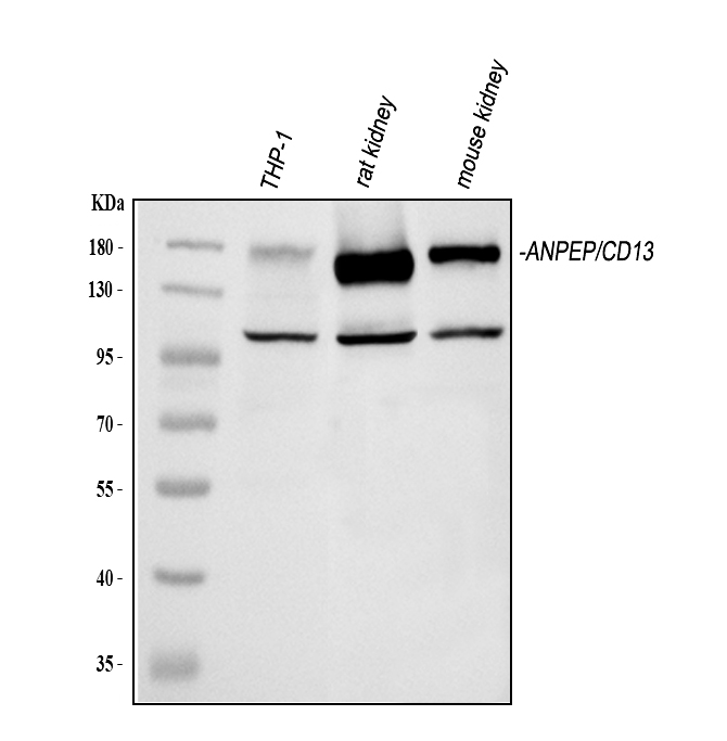
Western blot analysis of anti- CD13/Anpep antibody. The sample well of each lane was loaded with 30ug of sample under reducing conditions.Lane 1: human THP-1 whole cell lysates,Lane 2: rat kidney tissue lysates,Lane 3: mouse kidney tissue lysates.Use rabbit anti- CD13/Anpep 1:1000, probed with a goat anti-rabbit IgG-HRP secondary antibody. The signal is developed using an Enhanced Chemiluminescent detection (ECL) kit.IHC
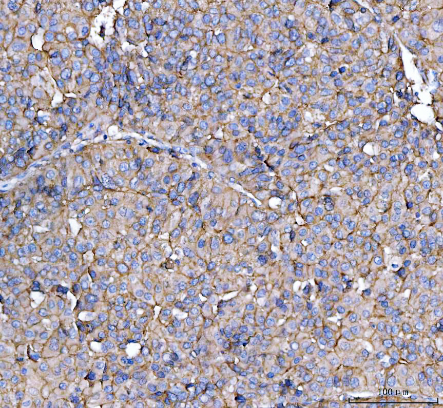
IHC analysis of CD13/ANPEP using anti-CD13/ANPEP antibody.CD13/ANPEP was detected in a paraffin-embedded section of human liver cancer tissue. The tissue section was incubated with rabbit anti-CD13/ANPEP Antibody at a dilution of 1:200 and developed using HRP Conjugated Rabbit IgG Super Vision Assay Kit with DAB as the chromogen.IHC
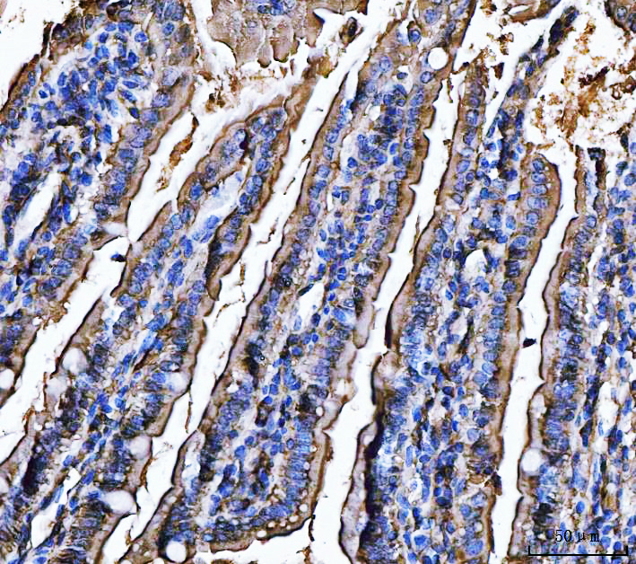
IHC analysis of CD13/ANPEP using anti-CD13/ANPEP antibody.CD13/ANPEP was detected in a paraffin-embedded section of mouse colon tissue. The tissue section was incubated with rabbit anti-CD13/ANPEP Antibody at a dilution of 1:200 and developed using HRP Conjugated Rabbit IgG Super Vision Assay Kit with DAB as the chromogen.IHC
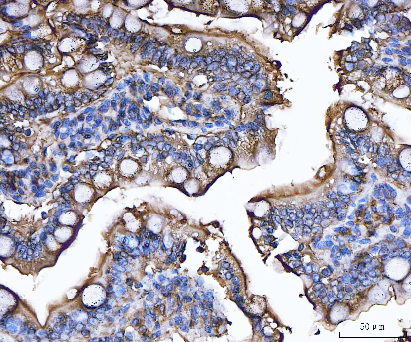
IHC analysis of CD13/ANPEP using anti-CD13/ANPEP antibody.CD13/ANPEP was detected in a paraffin-embedded section of rat colon tissue. The tissue section was incubated with rabbit anti-CD13/ANPEP Antibody at a dilution of 1:200 and developed using HRP Conjugated Rabbit IgG Super Vision Assay Kit with DAB as the chromogen.IF-P
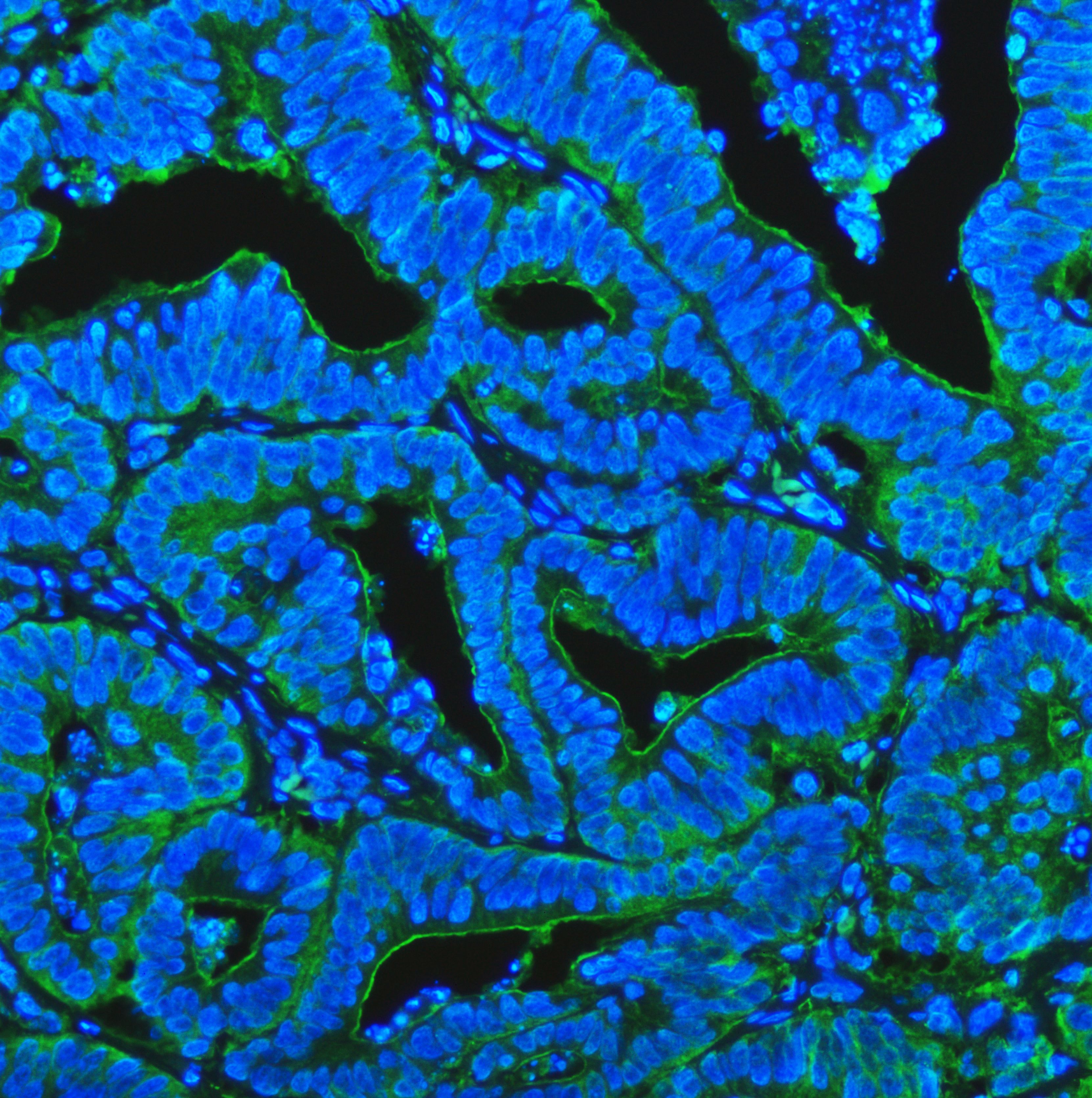
IF analysis using anti- CD13/Anpep antibody. detected in paraffin-embedded section of human colon cancer tissue. The tissue section were stained using the Fluoro488-conjugated Anti-rabbit IgG Secondary Antibody (green) and counterstained with DAPI (blue).IF-P
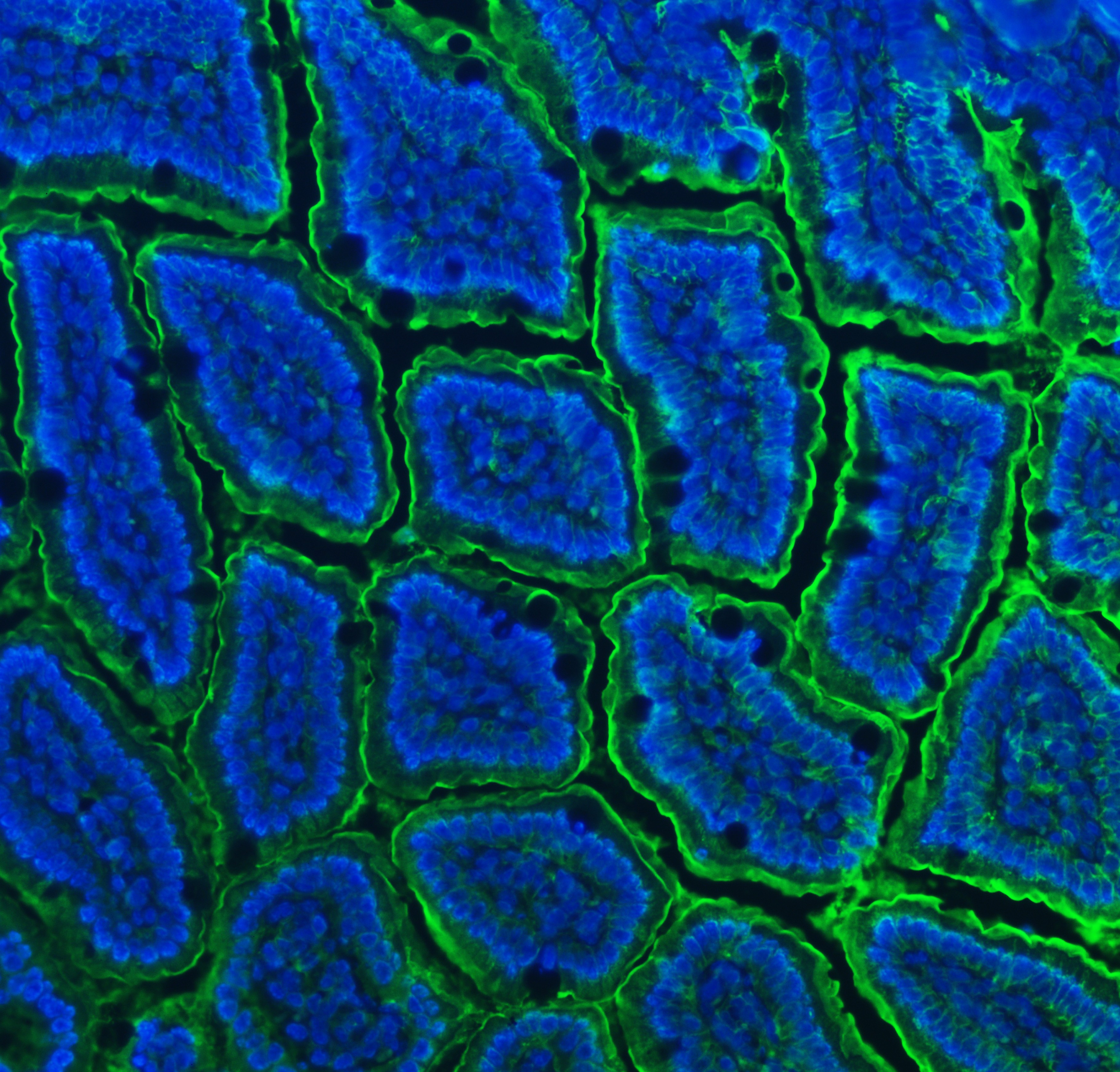
IF analysis using anti- CD13/Anpep antibody. detected in paraffin-embedded section of mouse colon tissue. The tissue section were stained using the Fluoro488-conjugated Anti-rabbit IgG Secondary Antibody (green) and counterstained with DAPI (blue).IF-P
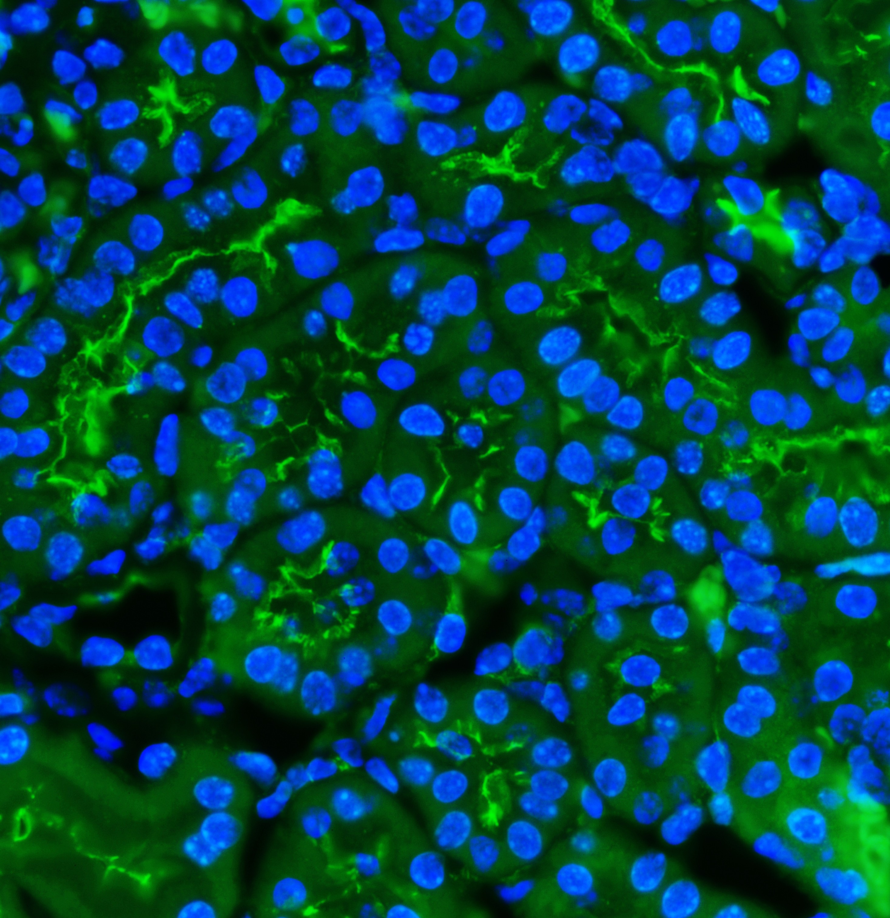
IF analysis using anti- CD13/Anpep antibody. detected in paraffin-embedded section of mouse kidney tissue. The tissue section were stained using the Fluoro488-conjugated Anti-rabbit IgG Secondary Antibody (green) and counterstained with DAPI (blue).IF-P
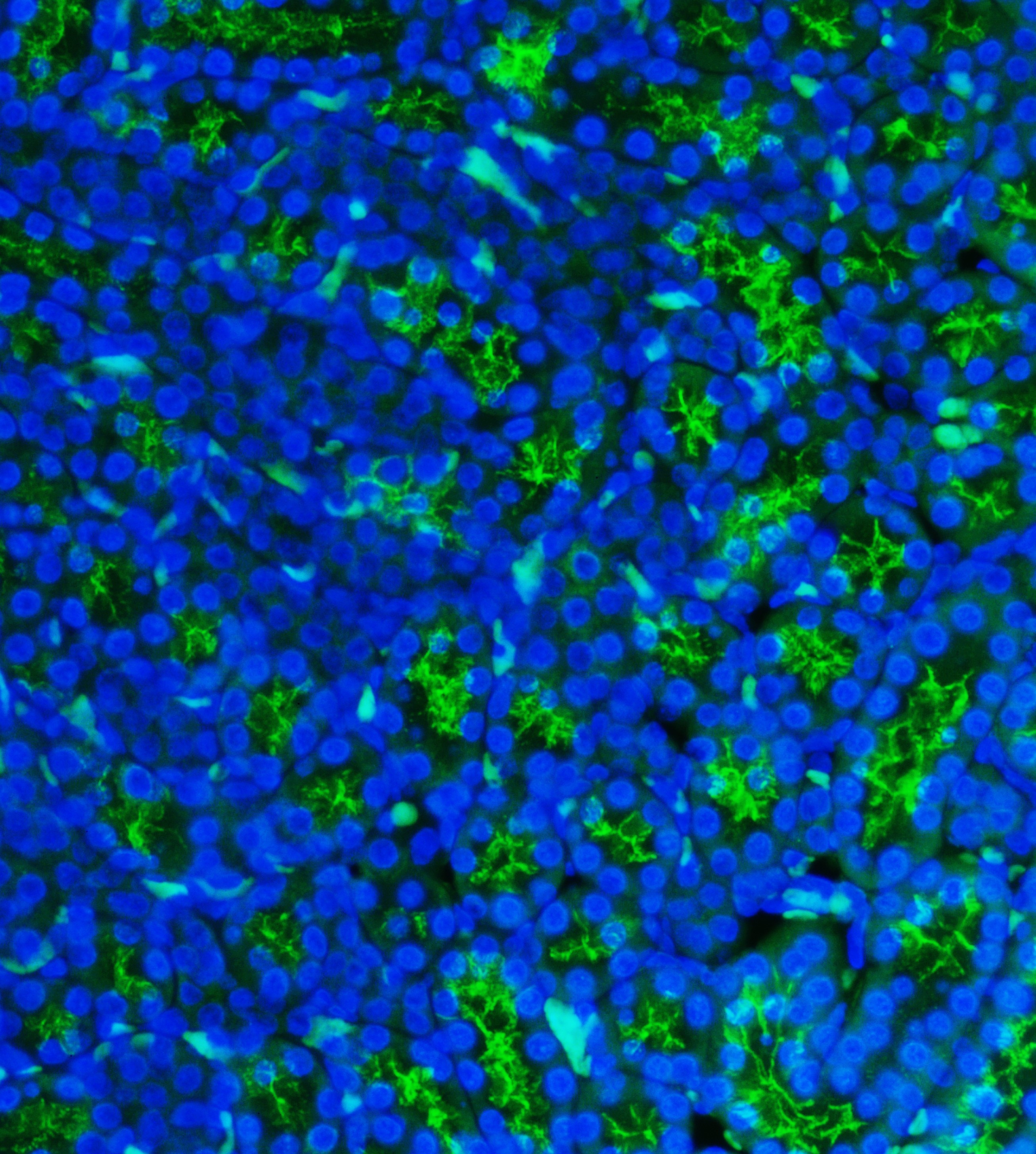
IF analysis using anti- CD13/Anpep antibody. detected in paraffin-embedded section of rat kidney tissue. The tissue section were stained using the Fluoro488-conjugated Anti-rabbit IgG Secondary Antibody (green) and counterstained with DAPI (blue).| Product Name | Rabbit polyclonal antibody to CD13/ANPEP |
|---|---|
| Antibody Type | Primary Antibodies |
| Immunogen | E.coli-derived rat CD13/Anpep recombinant protein (Position: Q642-S965) |
| Clonality | Polyclonal |
|---|---|
| Isotype | IgG |
| Host Species | Rabbit |
| Tested Applications | IF-PIHCWB |
| WB:1:500-1:2000 IHC:1:50-1:400 IF-P:1:50-1:400 |
|
| Species Reactivity | HumanMouseRat |
| Concentration | 0.5mg/ml |
| Purification | Affinity purified |
| Gene Symbol | ANPEP |
|---|---|
| Gene Synonyms | APN AP-M AP-N CD13 LAP1 P150 PEPN hAPN GP150 |
| Gene Full Name | alanyl aminopeptidase, membrane |
| Gene Summary | Aminopeptidase N is located in the small-intestinal and renal microvillar membrane, and also in other plasma membranes. In the small intestine aminopeptidase N plays a role in the final digestion of peptides generated from hydrolysis of proteins by gastric and pancreatic proteases. Its function in proximal tubular epithelial cells and other cell types is less clear. The large extracellular carboxyterminal domain contains a pentapeptide consensus sequence characteristic of members of the zinc-binding metalloproteinase superfamily. Sequence comparisons with known enzymes of this class showed that CD13 and aminopeptidase N are identical. The latter enzyme was thought to be involved in the metabolism of regulatory peptides by diverse cell types, including small intestinal and renal tubular epithelial cells, macrophages, granulocytes, and synaptic membranes from the CNS. This membrane-bound zinc metalloprotease is known to serve as a receptor for the HCoV-229E alphacoronavirus as well as other non-human coronaviruses. This gene has also been shown to promote angiogenesis, tumor growth, and metastasis and defects in this gene are associated with various types of leukemia and lymphoma. [provided by RefSeq, Apr 2020] |
| Alternative Names | Alanyl aminopeptidase Aminopeptidase M Aminopeptidase N ANPEP AP M AP N APN CD13 gp150 hAPN LAP1 Microsomal aminopeptidase p150 PEPN |
| Molecular Weight(MW) | 109 kDa(Observed band size: 150 kDa) |
| Cellular Localization | Cell membrane,Membrane |
WB
Western blot analysis of anti- CD13/Anpep antibody. The sample well of each lane was loaded with 30ug of sample under reducing conditions.Lane 1: human THP-1 whole cell lysates,Lane 2: rat kidney tissue lysates,Lane 3: mouse kidney tissue lysates.Use rabbit anti- CD13/Anpep 1:1000, probed with a goat anti-rabbit IgG-HRP secondary antibody. The signal is developed using an Enhanced Chemiluminescent detection (ECL) kit.
IHC
IHC analysis of CD13/ANPEP using anti-CD13/ANPEP antibody.CD13/ANPEP was detected in a paraffin-embedded section of human liver cancer tissue. The tissue section was incubated with rabbit anti-CD13/ANPEP Antibody at a dilution of 1:200 and developed using HRP Conjugated Rabbit IgG Super Vision Assay Kit with DAB as the chromogen.
IHC
IHC analysis of CD13/ANPEP using anti-CD13/ANPEP antibody.CD13/ANPEP was detected in a paraffin-embedded section of mouse colon tissue. The tissue section was incubated with rabbit anti-CD13/ANPEP Antibody at a dilution of 1:200 and developed using HRP Conjugated Rabbit IgG Super Vision Assay Kit with DAB as the chromogen.
IHC
IHC analysis of CD13/ANPEP using anti-CD13/ANPEP antibody.CD13/ANPEP was detected in a paraffin-embedded section of rat colon tissue. The tissue section was incubated with rabbit anti-CD13/ANPEP Antibody at a dilution of 1:200 and developed using HRP Conjugated Rabbit IgG Super Vision Assay Kit with DAB as the chromogen.
IF-P
IF analysis using anti- CD13/Anpep antibody. detected in paraffin-embedded section of human colon cancer tissue. The tissue section were stained using the Fluoro488-conjugated Anti-rabbit IgG Secondary Antibody (green) and counterstained with DAPI (blue).
IF-P
IF analysis using anti- CD13/Anpep antibody. detected in paraffin-embedded section of mouse colon tissue. The tissue section were stained using the Fluoro488-conjugated Anti-rabbit IgG Secondary Antibody (green) and counterstained with DAPI (blue).
IF-P
IF analysis using anti- CD13/Anpep antibody. detected in paraffin-embedded section of mouse kidney tissue. The tissue section were stained using the Fluoro488-conjugated Anti-rabbit IgG Secondary Antibody (green) and counterstained with DAPI (blue).
IF-P
IF analysis using anti- CD13/Anpep antibody. detected in paraffin-embedded section of rat kidney tissue. The tissue section were stained using the Fluoro488-conjugated Anti-rabbit IgG Secondary Antibody (green) and counterstained with DAPI (blue).| Application Notes | WB:1:500-1:2000 IHC:1:50-1:400 IF-P:1:50-1:400 |
|---|
| Form | Liquid |
|---|---|
| Storage Instructions | 12 months from date of receipt, -20℃ as supplied. 6 months 2 to 8℃ after reconstitution. Avoid repeated freezing and thawing. |
| Storage Buffer | 500ug/ml antibody with PBS, 0.02% NaN3, 1 mg/ml BSA and 50% glycerol. |


