IHC
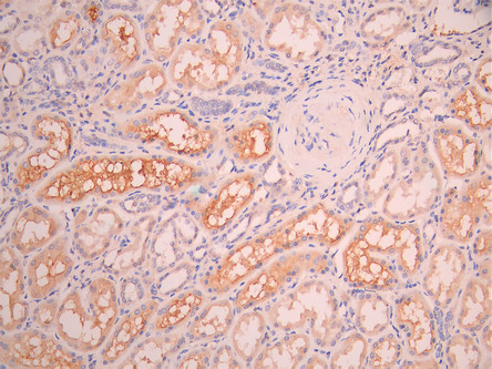
IHC image of ITGB8 Recombinant Monoclonal Antibody diluted at 1:50 and staining in paraffin-embedded human kidney tissue performed on a Leica BondTM system. After dewaxing and hydration, antigen retrieval was mediated by high pressure in a citrate buffer (pH 6.0). Section was blocked with 10% normal goat serum 30min at RT. Then primary antibody (1% BSA) was incubated at 4°C overnight. The primary is detected by a Anti-Human lgG, Fcy Fragment Specific labeled by HRP and visualized using 0.05% DAB.IHC
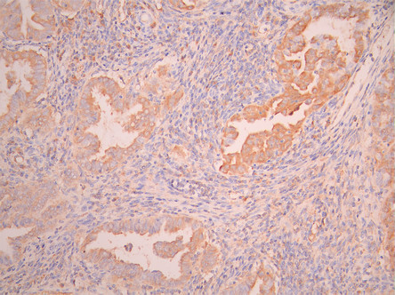
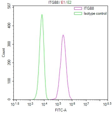
IHC image of ITGB8 Recombinant Monoclonal Antibody diluted at 1:50 and staining in paraffin-embedded human endometrial cancer performed on a Leica BondTM system. After dewaxing and hydration, antigen retrieval was mediated by high pressure in a citrate buffer (pH 6.0). Section was blocked with 10% normal goat serum 30min at RT. Then primary antibody (1% BSA) was incubated at 4°C overnight. The primary is detected by a Anti-Human lgG, Fcy Fragment Specific labeled by HRP and visualized using 0.05% DAB.Overlay Peak curve showing 786-O cells stained with ITGB8 Recombinant Monoclonal Antibody (red line) at 1:100. Then 10% normal goat serum to block non-specific protein-protein interactions followed by the antibody (1ug/1*106cells) for 45min at 4℃. The secondary antibody used was Fluorescein (FITC) AffiniPure Goat Anti-Human IgG, Fcγ fragment specific at 1:200 dilution for 35min at 4℃.Control antibody (green line) was human IgG1 (1ug/1*106cells) used under the same conditions. Acquisition of >10,000 events was performed.
| Product Name | ITGB8 Recombinant Monoclonal Antibody |
|---|---|
| Antibody Type | Primary Antibodies |
| Immunogen | Recombinant Human ITGB8 protein |
| Clonality | Monoclonal |
|---|---|
| Isotype | IgG1 |
| Host Species | Human |
| Tested Applications | FCIHC |
| IHC:1:50-1:200 FC:1:50-1:200 |
|
| Species Reactivity | Human |
| Concentration | 1mg/ml |
| Purification | Affinity purified |
| Gene Symbol | ITGB8 |
|---|---|
| Gene Full Name | integrin subunit beta 8 |
| Gene Summary | This gene is a member of the integrin beta chain family and encodes a single-pass type I membrane protein with a VWFA domain and four cysteine-rich repeats. This protein noncovalently binds to an alpha subunit to form a heterodimeric integrin complex. In general, integrin complexes mediate cell-cell and cell-extracellular matrix interactions and this complex plays a role in human airway epithelial proliferation. Alternatively spliced variants which encode different protein isoforms have been described; however, not all variants have been fully characterized. [provided by RefSeq, Jul 2008] |
| Alternative Names | Integrin beta-8 antibody ITB8_HUMAN antibody ITGB 8 antibody ITGB8 antibody |
| Cellular Localization | Cell membrane; Single-pass type I membrane protein. |
IHC
IHC image of ITGB8 Recombinant Monoclonal Antibody diluted at 1:50 and staining in paraffin-embedded human kidney tissue performed on a Leica BondTM system. After dewaxing and hydration, antigen retrieval was mediated by high pressure in a citrate buffer (pH 6.0). Section was blocked with 10% normal goat serum 30min at RT. Then primary antibody (1% BSA) was incubated at 4°C overnight. The primary is detected by a Anti-Human lgG, Fcy Fragment Specific labeled by HRP and visualized using 0.05% DAB.
IHC
IHC image of ITGB8 Recombinant Monoclonal Antibody diluted at 1:50 and staining in paraffin-embedded human endometrial cancer performed on a Leica BondTM system. After dewaxing and hydration, antigen retrieval was mediated by high pressure in a citrate buffer (pH 6.0). Section was blocked with 10% normal goat serum 30min at RT. Then primary antibody (1% BSA) was incubated at 4°C overnight. The primary is detected by a Anti-Human lgG, Fcy Fragment Specific labeled by HRP and visualized using 0.05% DAB.
Overlay Peak curve showing 786-O cells stained with ITGB8 Recombinant Monoclonal Antibody (red line) at 1:100. Then 10% normal goat serum to block non-specific protein-protein interactions followed by the antibody (1ug/1*106cells) for 45min at 4℃. The secondary antibody used was Fluorescein (FITC) AffiniPure Goat Anti-Human IgG, Fcγ fragment specific at 1:200 dilution for 35min at 4℃.Control antibody (green line) was human IgG1 (1ug/1*106cells) used under the same conditions. Acquisition of >10,000 events was performed.
| Application Notes | IHC:1:50-1:200 FC:1:50-1:200 |
|---|
| Form | Liquid |
|---|---|
| Storage Instructions | Upon receipt, store at -20°C or -80°C. Avoid repeated freeze. |
| Storage Buffer | 0.03% Proclin 300 Constituents: 50% Glycerol, 0.01M PBS, PH 7.4 |


