WB
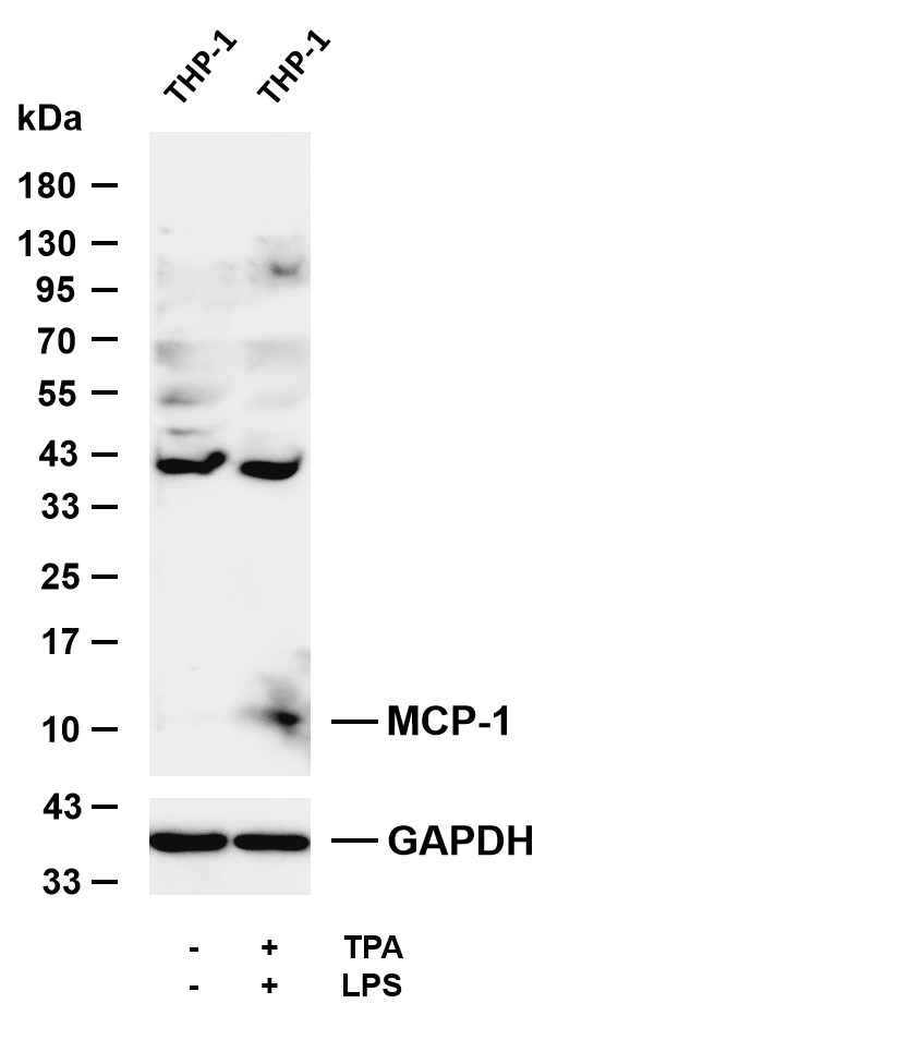
Various whole cell lysates were separated by 4-20% SDS-PAGE, and the membrane was blotted with anti-MCP-1 antibody. The HRP-conjugated Goat anti-Rabbit IgG (H + L) antibody was used to detect the antibody.WB
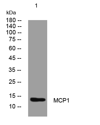
Western blot analysis of lysates from Hela cells, primary antibody was diluted at 1:1000, 4℃ over nightIHC
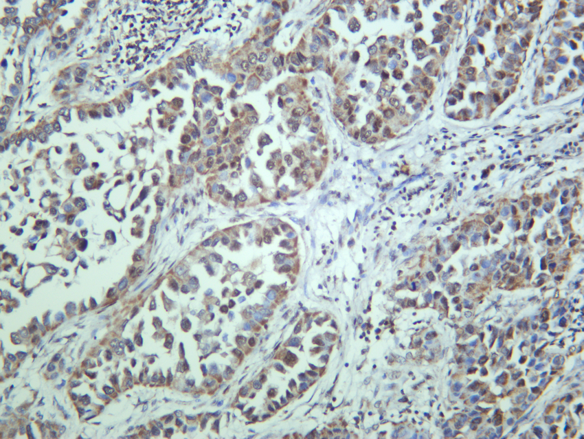
Human lung adenocarcinoma was stained with anti-MCP-1 rabbit antibodyIF/ICC
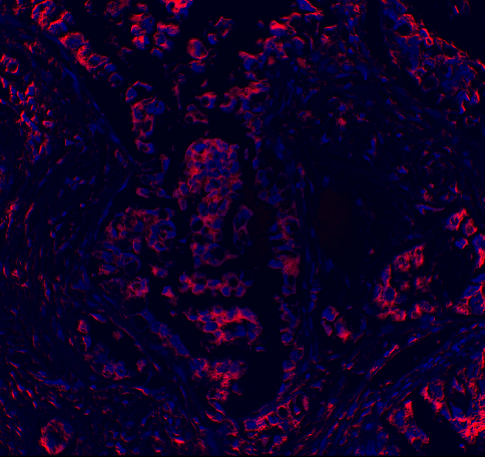
Immunofluorescence analysis of Human lung adenocarcinoma| Product Name | MCP-1 Rabbit mAb |
|---|---|
| Antibody Type | Primary Antibodies |
| Immunogen | The specific immunogen used to produce this antibody is proprietary information. |
| Clonality | Monoclonal |
|---|---|
| Isotype | IgG,K |
| Host Species | Rabbit |
| Tested Applications | ICC/IFIHCWB |
| IHC:1:2000-1:10000 WB:1:1000-1:5000 ICC/IF:1:200-1:1000 |
|
| Species Reactivity | Human |
| Concentration | 1mg/ml |
| Purification | Affinity purified |
| Gene Symbol | CCL2 |
|---|---|
| Gene Synonyms | HC11 MCAF MCP1 MCP-1 SCYA2 GDCF-2 SMC-CF HSMCR30 |
| Gene Full Name | C-C motif chemokine ligand 2 |
| Gene Summary | This gene is one of several cytokine genes clustered on the q-arm of chromosome 17. Chemokines are a superfamily of secreted proteins involved in immunoregulatory and inflammatory processes. The superfamily is divided into four subfamilies based on the arrangement of N-terminal cysteine residues of the mature peptide. This chemokine is a member of the CC subfamily which is characterized by two adjacent cysteine residues. This cytokine displays chemotactic activity for monocytes and basophils but not for neutrophils or eosinophils. It has been implicated in the pathogenesis of diseases characterized by monocytic infiltrates, like psoriasis, rheumatoid arthritis and atherosclerosis. It binds to chemokine receptors CCR2 and CCR4. Elevated expression of the encoded protein is associated with severe acute respiratory syndrome coronavirus 2 (SARS‐CoV‐2) infection. [provided by RefSeq, Aug 2020] |
| Molecular Weight(MW) | 11 kDa |
| Cellular Localization | Secreted |
WB
Various whole cell lysates were separated by 4-20% SDS-PAGE, and the membrane was blotted with anti-MCP-1 antibody. The HRP-conjugated Goat anti-Rabbit IgG (H + L) antibody was used to detect the antibody.
WB
Western blot analysis of lysates from Hela cells, primary antibody was diluted at 1:1000, 4℃ over night
IHC
Human lung adenocarcinoma was stained with anti-MCP-1 rabbit antibody
IF/ICC
Immunofluorescence analysis of Human lung adenocarcinoma| Application Notes | IHC:1:2000-1:10000 WB:1:1000-1:5000 ICC/IF:1:200-1:1000 |
|---|
| Form | Liquid |
|---|---|
| Storage Instructions | -15°C to -25°C/1 year(Do not lower than -25°C) |
| Storage Buffer | PBS, 50% glycerol, 0.05% Proclin 300, 0.05%BSA |


