WB
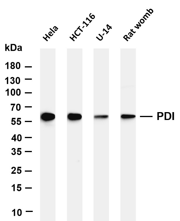
Various whole cell lysates were separated by 4-20% SDS-PAGE, and the membrane was blotted with anti-PDI antibody. The HRP-conjugated Goat anti-Rabbit IgG (H + L) antibody was used to detect the antibody.IHC
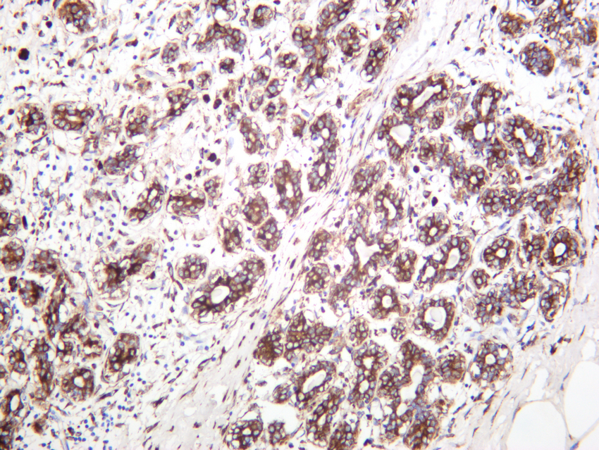
Human breast carcinoma was stained with anti-PDI rabbit antibody.IHC
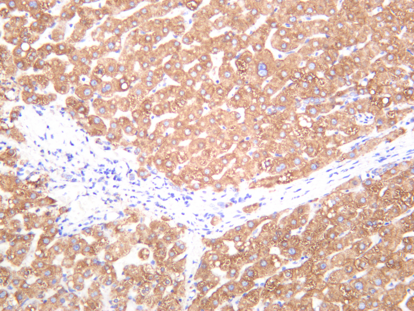
Human liver was stained with anti-PDI rabbit antibody.IHC
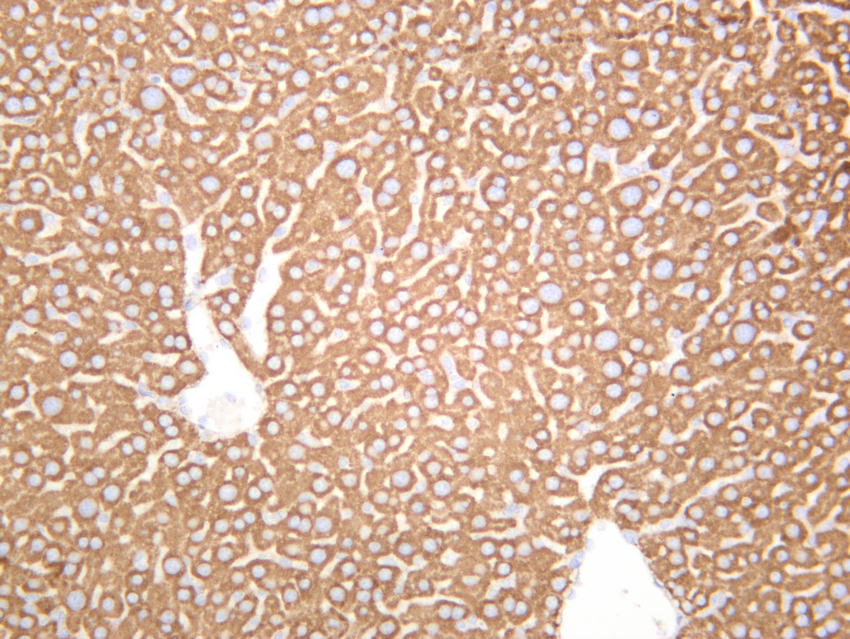
Rat liver was stained with anti-PDI rabbit antibody.ICC/IF
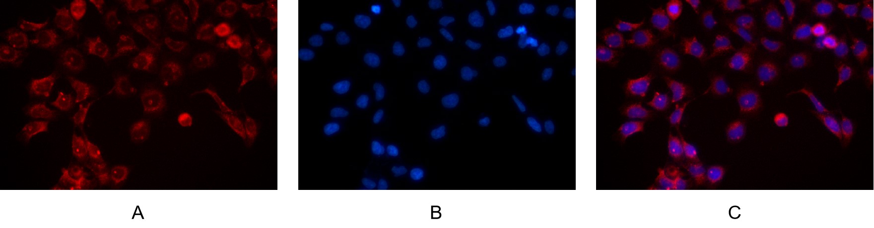
Immunofluorescence analysis of HEK293. Picture A: P4HB antibody (red). Picture B: DAPI (blue). Picture C: Merge of A+B| Product Name | PDI Rabbit mAb |
|---|---|
| Antibody Type | Primary Antibodies |
| Immunogen | The specific immunogen used to produce this antibody is proprietary information. |
| Clonality | Monoclonal |
|---|---|
| Isotype | IgG,K |
| Host Species | Rabbit |
| Tested Applications | ICC/IFIHCWB |
| IHC:1:200-1:1000 WB:1:2000-1:10000 ICC/IF:1:200-1:1000 |
|
| Species Reactivity | HumanMouseRat |
| Concentration | 1mg/ml |
| Purification | Protein A |
| Gene Symbol | P4HB |
|---|---|
| Gene Synonyms | DSI GIT PDI PHDB PDIA1 PO4DB PO4HB PROHB CLCRP1 ERBA2L P4Hbeta |
| Gene Full Name | prolyl 4-hydroxylase subunit beta |
| Gene Summary | This gene encodes the beta subunit of prolyl 4-hydroxylase, a highly abundant multifunctional enzyme that belongs to the protein disulfide isomerase family. When present as a tetramer consisting of two alpha and two beta subunits, this enzyme is involved in hydroxylation of prolyl residues in preprocollagen. This enzyme is also a disulfide isomerase containing two thioredoxin domains that catalyze the formation, breakage and rearrangement of disulfide bonds. Other known functions include its ability to act as a chaperone that inhibits aggregation of misfolded proteins in a concentration-dependent manner, its ability to bind thyroid hormone, its role in both the influx and efflux of S-nitrosothiol-bound nitric oxide, and its function as a subunit of the microsomal triglyceride transfer protein complex. [provided by RefSeq, Jul 2008] |
| Molecular Weight(MW) | 57 kDa |
| Cellular Localization | Endoplasmic reticulum |
WB
Various whole cell lysates were separated by 4-20% SDS-PAGE, and the membrane was blotted with anti-PDI antibody. The HRP-conjugated Goat anti-Rabbit IgG (H + L) antibody was used to detect the antibody.
IHC
Human breast carcinoma was stained with anti-PDI rabbit antibody.
IHC
Human liver was stained with anti-PDI rabbit antibody.
IHC
Rat liver was stained with anti-PDI rabbit antibody.
ICC/IF
Immunofluorescence analysis of HEK293. Picture A: P4HB antibody (red). Picture B: DAPI (blue). Picture C: Merge of A+B| Application Notes | IHC:1:200-1:1000 WB:1:2000-1:10000 ICC/IF:1:200-1:1000 |
|---|
| Form | Liquid |
|---|---|
| Storage Instructions | -15°C to -25°C/1 year(Do not lower than -25°C) |
| Storage Buffer | PBS, 50% glycerol, 0.05% Proclin 300, 0.05%BSA |


