WB
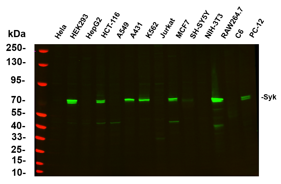
Various whole cell lysates were separated by 4-20% SDS-PAGE, and the primary antibody was used at 4℃ over night with a 1:5000 dilution . The Dylight 800-conjugated Goat anti-Rabbit antibody was used to detect the antibody.WB
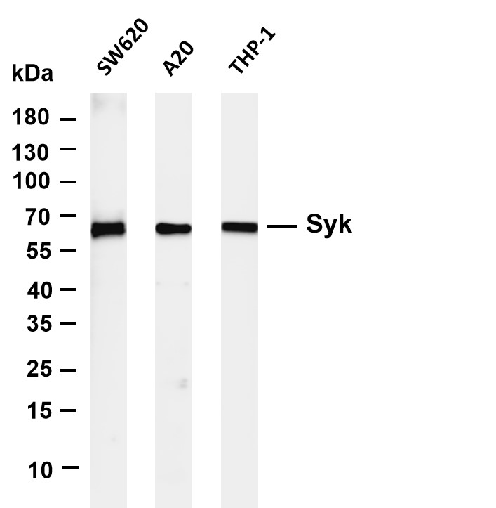
Various whole cell lysates were separated by 4-20% SDS-PAGE, and the membrane was blotted with anti-Syk antibody. The HRP-conjugated Goat anti-Rabbit IgG (H + L) antibody was used to detect the antibody.IHC
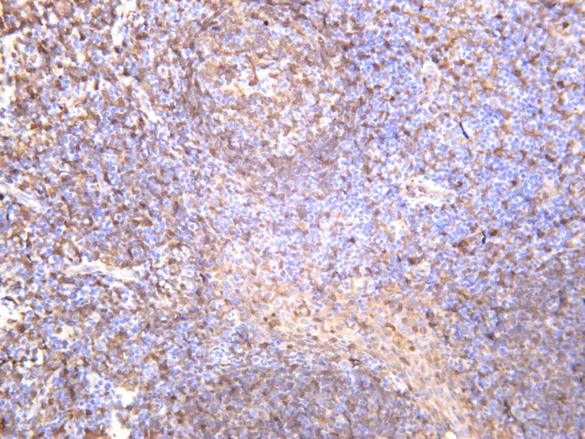
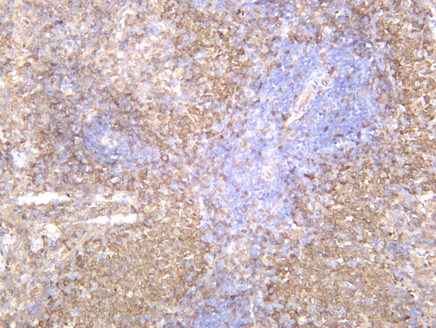
Human spleen was stained with anti-Syk rabbit antibody.IHC
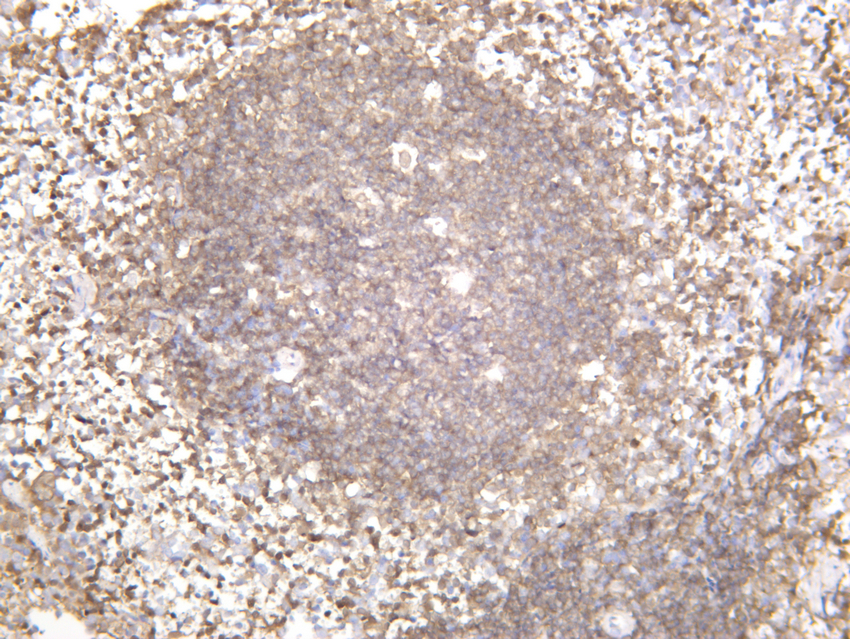
Mouse spleen was stained with anti-Syk rabbit antibody.IHC
Rat spleen was stained with anti-Syk rabbit antibody.ICC/IF
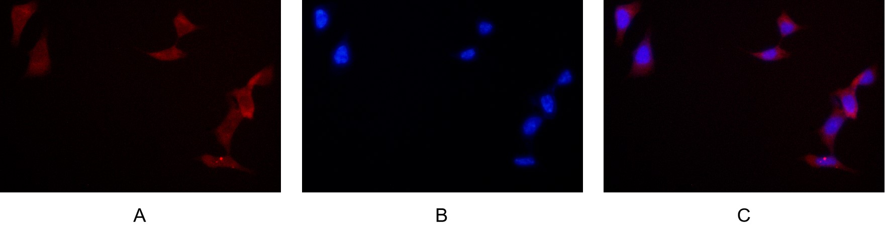
Immunofluorescence analysis of HEK293. Picture A: Syk antibody (red). Picture B: DAPI (blue). Picture C: Merge of A+B.| Product Name | Syk Rabbit mAb |
|---|---|
| Antibody Type | Primary Antibodies |
| Immunogen | Synthesized peptide derived from the N-terminal region of human Syk. |
| Clonality | Monoclonal |
|---|---|
| Isotype | IgG,K |
| Host Species | Rabbit |
| Tested Applications | ICC/IFIHCWB |
| IHC:1:400-1:1000 WB:1:2000-1:10000 ICC/IF:1:200-1:1000 |
|
| Species Reactivity | HumanMouseRat |
| Concentration | 1mg/ml |
| Purification | Protein A |
| Gene Symbol | SYK |
|---|---|
| Gene Synonyms | IMD82 p72-Syk |
| Gene Full Name | spleen associated tyrosine kinase |
| Gene Summary | This gene encodes a member of the family of non-receptor type Tyr protein kinases. This protein is widely expressed in hematopoietic cells and is involved in coupling activated immunoreceptors to downstream signaling events that mediate diverse cellular responses, including proliferation, differentiation, and phagocytosis. It is thought to be a modulator of epithelial cell growth and a potential tumour suppressor in human breast carcinomas. Alternatively spliced transcript variants encoding different isoforms have been found for this gene. [provided by RefSeq, Mar 2010] |
| Alternative Names | SYK Tyrosine-protein kinase SYK Spleen tyrosine kinase p72-Syk |
| Molecular Weight(MW) | 72 kDa (Observed band size: 65kDa) |
| Cellular Localization | Membrane. |
WB
Various whole cell lysates were separated by 4-20% SDS-PAGE, and the primary antibody was used at 4℃ over night with a 1:5000 dilution . The Dylight 800-conjugated Goat anti-Rabbit antibody was used to detect the antibody.
WB
Various whole cell lysates were separated by 4-20% SDS-PAGE, and the membrane was blotted with anti-Syk antibody. The HRP-conjugated Goat anti-Rabbit IgG (H + L) antibody was used to detect the antibody.
IHC
Human spleen was stained with anti-Syk rabbit antibody.
IHC
Mouse spleen was stained with anti-Syk rabbit antibody.
IHC
Rat spleen was stained with anti-Syk rabbit antibody.
ICC/IF
Immunofluorescence analysis of HEK293. Picture A: Syk antibody (red). Picture B: DAPI (blue). Picture C: Merge of A+B.| Application Notes | IHC:1:400-1:1000 WB:1:2000-1:10000 ICC/IF:1:200-1:1000 |
|---|
| Form | Liquid |
|---|---|
| Storage Instructions | -15°C to -25°C/1 year(Do not lower than -25°C) |
| Storage Buffer | PBS, 50% glycerol, 0.05% Proclin 300, 0.05%BSA |


