WB
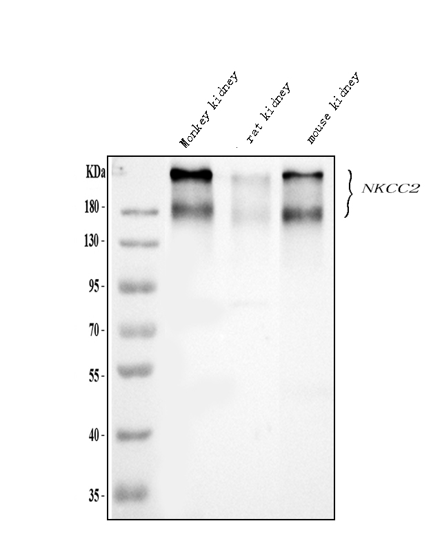
Western blot analysis of NKCC2/SLC12A1 using anti-NKCC2/SLC12A1 antibody. The sample well of each lane was loaded with 30 ug of sample under reducing conditions. After electrophoresis, proteins were transferred to a membrane. Then the membrane was incubated with rabbit anti-NKCC2/SLC12A1 antigen affinity purified polyclonal antibody at a dilution of 1:1000 and probed with a goat anti-rabbit IgG-HRP secondary antibody. The signal is developed using ECL Plus Western Blotting Substrate.IHC
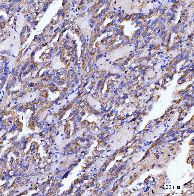
IHC analysis of NKCC2/SLC12A1 using anti-NKCC2/SLC12A1 antibody. NKCC2/SLC12A1 was detected in a paraffin-embedded section of human renal cancer tissue. Biotinylated goat anti-rabbit IgG was used as secondary antibody. The tissue section was incubated with rabbit anti-NKCC2/SLC12A1 Antibody at a dilution of 1:200 and developed using Strepavidin-Biotin-Complex (SABC) with DAB as the chromogen.IHC
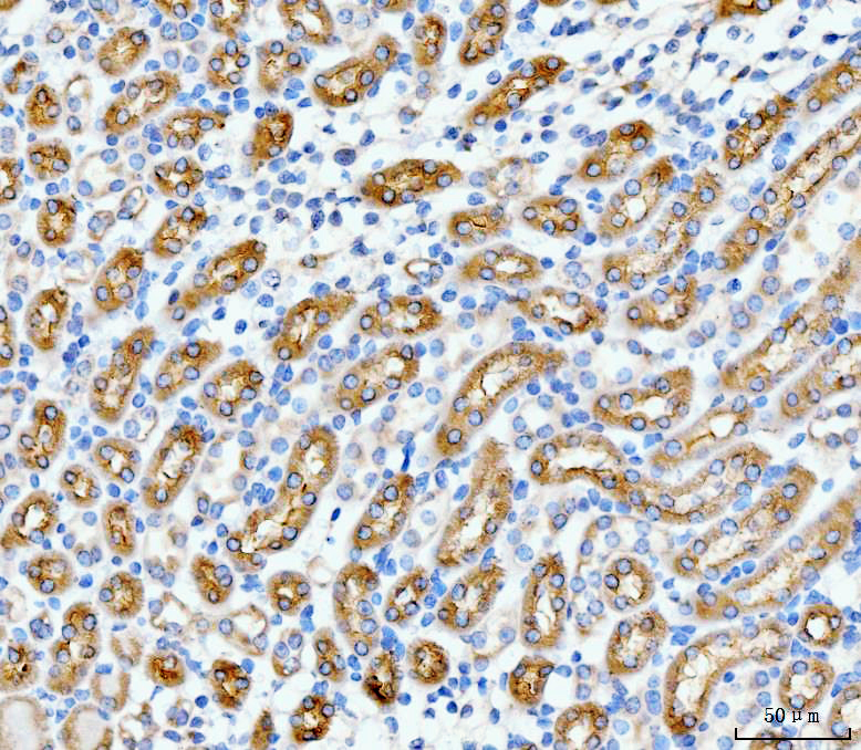
IHC analysis of NKCC2/SLC12A1 using anti-NKCC2/SLC12A1 antibody. NKCC2/SLC12A1 was detected in a paraffin-embedded section of mouse kidney tissue. Biotinylated goat anti-rabbit IgG was used as secondary antibody. The tissue section was incubated with rabbit anti-NKCC2/SLC12A1 Antibody at a dilution of 1:200 and developed using Strepavidin-Biotin-Complex (SABC) with DAB as the chromogen.IHC
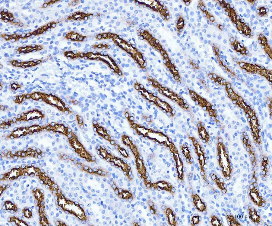
IHC analysis of NKCC2/SLC12A1 using anti-NKCC2/SLC12A1 antibody. NKCC2/SLC12A1 was detected in a paraffin-embedded section of rat kidney tissue. Biotinylated goat anti-rabbit IgG was used as secondary antibody. The tissue section was incubated with rabbit anti-NKCC2/SLC12A1 Antibody at a dilution of 1:200 and developed using Strepavidin-Biotin-Complex (SABC) with DAB as the chromogen.IF-P
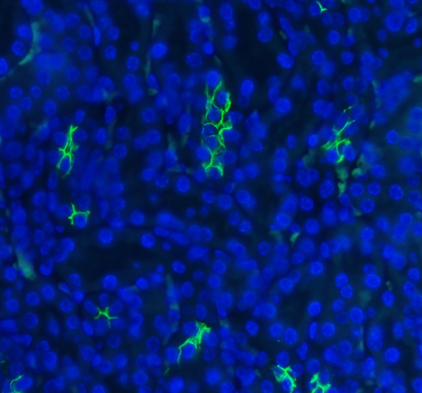
IF analysis of NKCC2/SLC12A1 using anti-NKCC2/SLC12A1 antibody. NKCC2/SLC12A1 was detected in a paraffin-embedded section of mouse kidney tissue. The tissue section was incubated with rabbit anti-NKCC2/SLC12A1 Antibody at a dilution of 1:100. Fluoro488 Conjugated Goat Anti-Rabbit IgG (Green) was used as secondary antibody. The section was counterstained with DAPI (Blue).IF-P
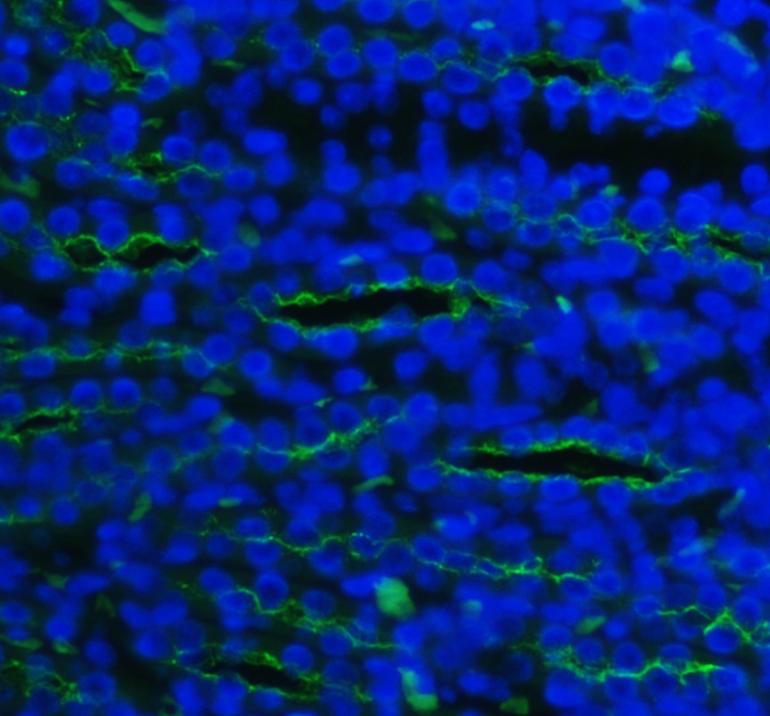
IF analysis of NKCC2/SLC12A1 using anti-NKCC2/SLC12A1 antibody. NKCC2/SLC12A1 was detected in a paraffin-embedded section of rat kidney tissue. The tissue section was incubated with rabbit anti-NKCC2/SLC12A1 Antibody at a dilution of 1:100. Fluoro488 Conjugated Goat Anti-Rabbit IgG (Green) was used as secondary antibody. The section was counterstained with DAPI (Blue).| Product Name | Rabbit polyclonal antibody to NKCC2/SLC12A1 |
|---|---|
| Antibody Type | Primary Antibodies |
| Immunogen | A synthetic peptide corresponding to a sequence at the N-terminus of human SLC12A1, different from the related mouse sequence by two amino acids, and from the related rat sequence by four amino acids. |
| Clonality | Polyclonal |
|---|---|
| Isotype | IgG |
| Host Species | Rabbit |
| Tested Applications | IF-PIHCWB |
| WB:1:500-1:2000 IHC:1:50-1:400 IF-P:1:50-1:400 |
|
| Species Reactivity | HumanMonkeyMouseRat |
| Concentration | 0.5mg/ml |
| Purification | Affinity purified |
| Gene Symbol | SLC12A1 |
|---|---|
| Gene Synonyms | BSC BSC1 CCC2 BSC-1 NKCC2 |
| Gene Full Name | solute carrier family 12 member 1 |
| Gene Summary | This gene encodes a kidney-specific sodium-potassium-chloride cotransporter that is expressed on the luminal membrane of renal epithelial cells of the thick ascending limb of Henle's loop and the macula densa. It plays a key role in concentrating urine and accounts for most of the NaCl resorption. It is sensitive to such diuretics as furosemide and bumetanide. Some Bartter-like syndromes result from defects in this gene. Alternative splicing results in multiple transcript variants encoding distinct isoforms. Additional splice variants have been described but their biological validity in humans has not been experimentally proven.[provided by RefSeq, May 2010] |
| Alternative Names | BSC1 NKCC2 SLC12A1 |
| Molecular Weight(MW) | 121 kDa(Observed band size: 150-290 kDa) |
| Cellular Localization | Cell membrane,Membrane |
WB
Western blot analysis of NKCC2/SLC12A1 using anti-NKCC2/SLC12A1 antibody. The sample well of each lane was loaded with 30 ug of sample under reducing conditions. After electrophoresis, proteins were transferred to a membrane. Then the membrane was incubated with rabbit anti-NKCC2/SLC12A1 antigen affinity purified polyclonal antibody at a dilution of 1:1000 and probed with a goat anti-rabbit IgG-HRP secondary antibody. The signal is developed using ECL Plus Western Blotting Substrate.
IHC
IHC analysis of NKCC2/SLC12A1 using anti-NKCC2/SLC12A1 antibody. NKCC2/SLC12A1 was detected in a paraffin-embedded section of human renal cancer tissue. Biotinylated goat anti-rabbit IgG was used as secondary antibody. The tissue section was incubated with rabbit anti-NKCC2/SLC12A1 Antibody at a dilution of 1:200 and developed using Strepavidin-Biotin-Complex (SABC) with DAB as the chromogen.
IHC
IHC analysis of NKCC2/SLC12A1 using anti-NKCC2/SLC12A1 antibody. NKCC2/SLC12A1 was detected in a paraffin-embedded section of mouse kidney tissue. Biotinylated goat anti-rabbit IgG was used as secondary antibody. The tissue section was incubated with rabbit anti-NKCC2/SLC12A1 Antibody at a dilution of 1:200 and developed using Strepavidin-Biotin-Complex (SABC) with DAB as the chromogen.
IHC
IHC analysis of NKCC2/SLC12A1 using anti-NKCC2/SLC12A1 antibody. NKCC2/SLC12A1 was detected in a paraffin-embedded section of rat kidney tissue. Biotinylated goat anti-rabbit IgG was used as secondary antibody. The tissue section was incubated with rabbit anti-NKCC2/SLC12A1 Antibody at a dilution of 1:200 and developed using Strepavidin-Biotin-Complex (SABC) with DAB as the chromogen.
IF-P
IF analysis of NKCC2/SLC12A1 using anti-NKCC2/SLC12A1 antibody. NKCC2/SLC12A1 was detected in a paraffin-embedded section of mouse kidney tissue. The tissue section was incubated with rabbit anti-NKCC2/SLC12A1 Antibody at a dilution of 1:100. Fluoro488 Conjugated Goat Anti-Rabbit IgG (Green) was used as secondary antibody. The section was counterstained with DAPI (Blue).
IF-P
IF analysis of NKCC2/SLC12A1 using anti-NKCC2/SLC12A1 antibody. NKCC2/SLC12A1 was detected in a paraffin-embedded section of rat kidney tissue. The tissue section was incubated with rabbit anti-NKCC2/SLC12A1 Antibody at a dilution of 1:100. Fluoro488 Conjugated Goat Anti-Rabbit IgG (Green) was used as secondary antibody. The section was counterstained with DAPI (Blue).| Application Notes | WB:1:500-1:2000 IHC:1:50-1:400 IF-P:1:50-1:400 |
|---|
| Form | Liquid |
|---|---|
| Storage Instructions | 12 months from date of receipt, -20℃ as supplied. 6 months 2 to 8℃ after reconstitution. Avoid repeated freezing and thawing. |
| Storage Buffer | 500ug/ml antibody with PBS, 0.02% NaN3, 1 mg/ml BSA and 50% glycerol. |


