WB
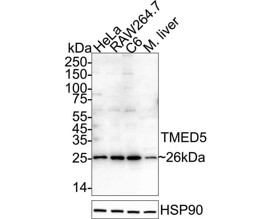
Western blot analysis of TMED5 on different lysates with Rabbit anti-TMED5 antibody at 1/100,000 dilution. Exposure time: 25 seconds; 4-20% SDS-PAGE gel. Proteins were transferred to a PVDF membrane and blocked with 5% NFDM/TBST for 1 hour at room temperature. The primary antibody at 1/100,000 dilution was used in primary antibody dilution at 4℃ overnight. Goat Anti-Rabbit IgG - HRP Secondary Antibody at 1/50,000 dilution was used for 1 hour at room temperature.IHC
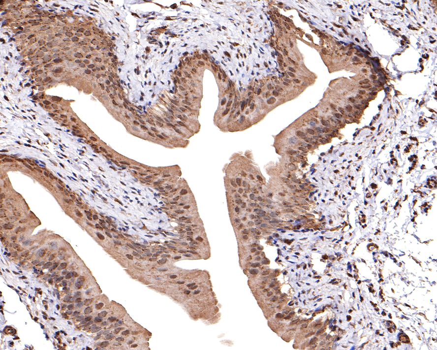
Immunohistochemical analysis of paraffin-embedded rat bladder tissue with Rabbit anti-TMED5 antibody at 1/600 dilution. The section was pre-treated using heat mediated antigen retrieval with sodium citrate buffer (pH 6.0) (high pressure) for 2 minutes. The tissues were blocked in 1% BSA for 20 minutes at room temperature, washed with ddH2O and PBS, and then probed with the primary antibody at 1/600 dilution for 1 hour at room temperature. The detection was performed using an HRP conjugated compact polymer system. DAB was used as the chromogen. Tissues were counterstained with hematoxylin and mounted with DPX.IHC
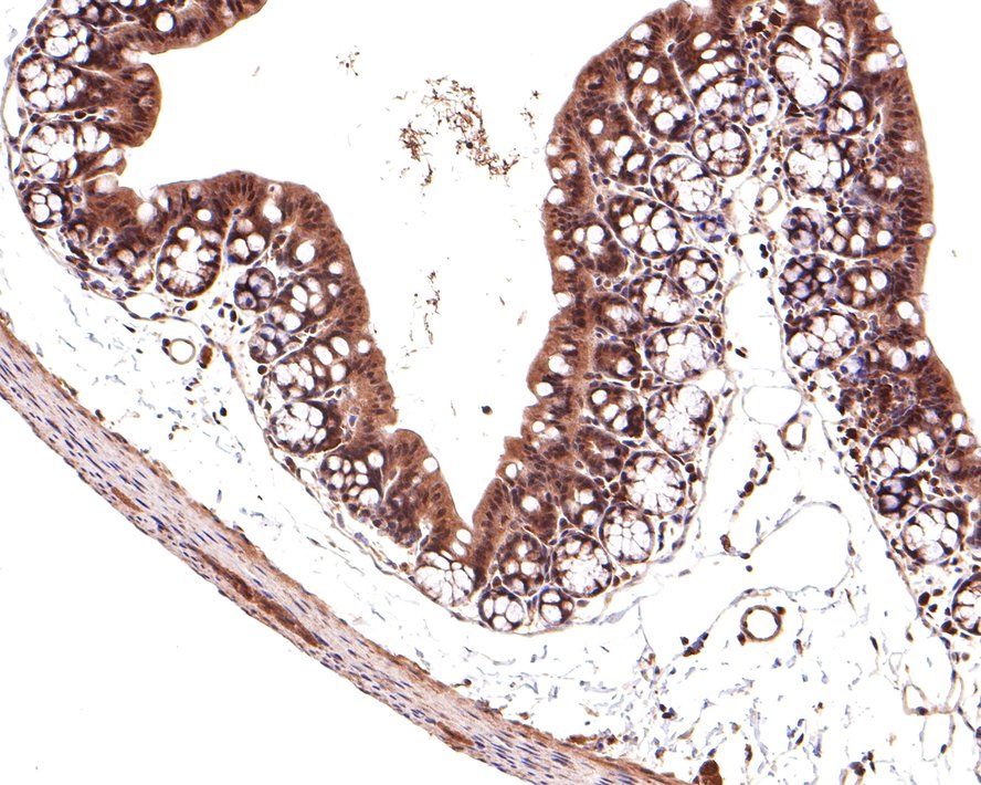
Immunohistochemical analysis of paraffin-embedded mouse large intestine tissue with Rabbit anti-TMED5 antibody at 1/5,000 dilution. The section was pre-treated using heat mediated antigen retrieval with sodium citrate buffer (pH 6.0) (high pressure) for 2 minutes. The tissues were blocked in 1% BSA for 20 minutes at room temperature, washed with ddH2O and PBS, and then probed with the primary antibody at 1/5,000 dilution for 1 hour at room temperature. The detection was performed using an HRP conjugated compact polymer system. DAB was used as the chromogen. Tissues were counterstained with hematoxylin and mounted with DPX.| Product Name | TMED5 Rabbit Polyclonal Antibody |
|---|---|
| Antibody Type | Primary Antibodies |
| Immunogen | Recombinant protein within Human TMED5 aa 1-200 / 229. |
| Clonality | Polyclonal |
|---|---|
| Isotype | IgG |
| Host Species | Rabbit |
| Tested Applications | IHCWB |
| WB:1:100000 IHC:1:600-1:5000 |
|
| Species Reactivity | HumanMouseRat |
| Concentration | 1mg/ml |
| Purification | Affinity purified |
| Gene Symbol | TMED5 |
|---|---|
| Gene Synonyms | p28 p24g2 CGI-100 |
| Gene Full Name | transmembrane p24 trafficking protein 5 |
| Gene Summary | Involved in Golgi ribbon formation. Located in cis-Golgi network; endoplasmic reticulum exit site; and endoplasmic reticulum-Golgi intermediate compartment. [provided by Alliance of Genome Resources, Jul 2025] |
| Alternative Names | CGI 100 antibody DKFZp781B2248 antibody p24 family protein gamma-2 antibody p24gamma2 antibody p28 antibody RP5 976O13.2 antibody TMED 5 antibody TMED5 antibody TMED5_HUMAN antibody Transmembrane emp24 domain containing protein 5 antibody CGI 100 antibody DKFZp781B2248 antibody p24 family protein gamma-2 antibody p24gamma2 antibody p28 antibody RP5 976O13.2 antibody TMED 5 antibody TMED5 antibody TMED5_HUMAN antibody Transmembrane emp24 domain containing protein 5 antibody Transmembrane emp24 domain-containing protein 5 antibody Transmembrane emp24 protein transport domain containing 5 antibody UNQ397/PRO733 antibody |
| Molecular Weight(MW) | 26 kDa |
| Cellular Localization | Cis-Golgi network membrane, endoplasmic reticulum membrane, endoplasmic reticulum-Golgi intermediate compartment membrane. |
WB
Western blot analysis of TMED5 on different lysates with Rabbit anti-TMED5 antibody at 1/100,000 dilution. Exposure time: 25 seconds; 4-20% SDS-PAGE gel. Proteins were transferred to a PVDF membrane and blocked with 5% NFDM/TBST for 1 hour at room temperature. The primary antibody at 1/100,000 dilution was used in primary antibody dilution at 4℃ overnight. Goat Anti-Rabbit IgG - HRP Secondary Antibody at 1/50,000 dilution was used for 1 hour at room temperature.
IHC
Immunohistochemical analysis of paraffin-embedded rat bladder tissue with Rabbit anti-TMED5 antibody at 1/600 dilution. The section was pre-treated using heat mediated antigen retrieval with sodium citrate buffer (pH 6.0) (high pressure) for 2 minutes. The tissues were blocked in 1% BSA for 20 minutes at room temperature, washed with ddH2O and PBS, and then probed with the primary antibody at 1/600 dilution for 1 hour at room temperature. The detection was performed using an HRP conjugated compact polymer system. DAB was used as the chromogen. Tissues were counterstained with hematoxylin and mounted with DPX.
IHC
Immunohistochemical analysis of paraffin-embedded mouse large intestine tissue with Rabbit anti-TMED5 antibody at 1/5,000 dilution. The section was pre-treated using heat mediated antigen retrieval with sodium citrate buffer (pH 6.0) (high pressure) for 2 minutes. The tissues were blocked in 1% BSA for 20 minutes at room temperature, washed with ddH2O and PBS, and then probed with the primary antibody at 1/5,000 dilution for 1 hour at room temperature. The detection was performed using an HRP conjugated compact polymer system. DAB was used as the chromogen. Tissues were counterstained with hematoxylin and mounted with DPX.| Application Notes | WB:1:100000 IHC:1:600-1:5000 |
|---|
| Form | Liquid |
|---|---|
| Storage Instructions | Shipped at 4℃. Store at +4℃ short term (1-2 weeks). It is recommended to aliquot into single-use upon delivery. Store at -20℃ long term. |
| Storage Buffer | PBS (pH7.4), 0.1% BSA, 40% Glycerol. Preservative: 0.05% Sodium Azide. |


