WB
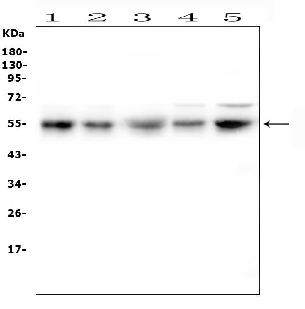
Western blot analysis of RBMS3 using anti-RBMS3 antibody. The sample well of each lane was loaded with 30 ug of sample under reducing conditions. Lane 1: human U2OS whole cell lysates, Lane 2: human PC-3 whole cell lysates, Lane 3: human U-87MG whole cell lysates, Lane 4: human A549 whole cell lysates, Lane 5: human Hela whole cell lysates. After electrophoresis, proteins were transferred to a membrane. Then the membrane was incubated with rabbit anti-RBMS3 antigen affinity purified polyclonal antibod at a dilution of 1:1000 and probed with a goat anti-rabbit IgG-HRP secondary antibody. The signal is developed using ECL Plus Western Blotting Substrate.IHC
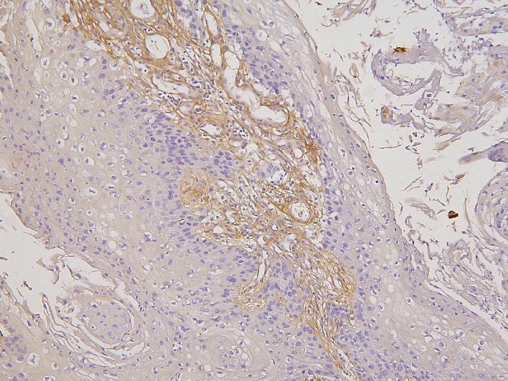
IHC analysis of RBMS3 using anti-RBMS3 antibody. RBMS3 was detected in a paraffin-embedded section of human oesophagus squama cancer tissue. Biotinylated goat anti-rabbit IgG was used as secondary antibody. The tissue section was incubated with rabbit anti-RBMS3 Antibody at a dilution of 1:200 and developed using Strepavidin-Biotin-Complex (SABC) with DAB as the chromogen.IHC
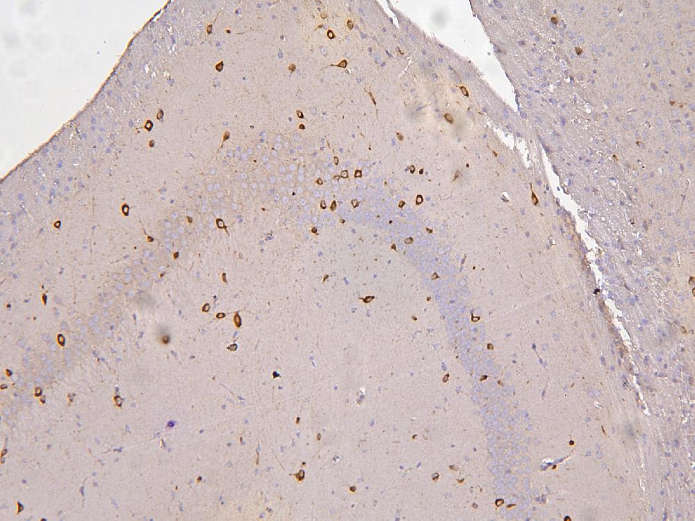
IHC analysis of RBMS3 using anti-RBMS3 antibody. RBMS3 was detected in a paraffin-embedded section of mouse brain tissue. Biotinylated goat anti-rabbit IgG was used as secondary antibody. The tissue section was incubated with rabbit anti-RBMS3 Antibody at a dilution of 1:200 and developed using Strepavidin-Biotin-Complex (SABC) with DAB as the chromogen.IHC
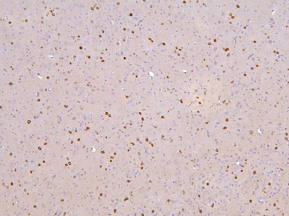
IHC analysis of RBMS3 using anti-RBMS3 antibody. RBMS3 was detected in a paraffin-embedded section of rat brain tissue. Biotinylated goat anti-rabbit IgG was used as secondary antibody. The tissue section was incubated with rabbit anti-RBMS3 Antibody at a dilution of 1:200 and developed using Strepavidin-Biotin-Complex (SABC) with DAB as the chromogen.ICC/IF
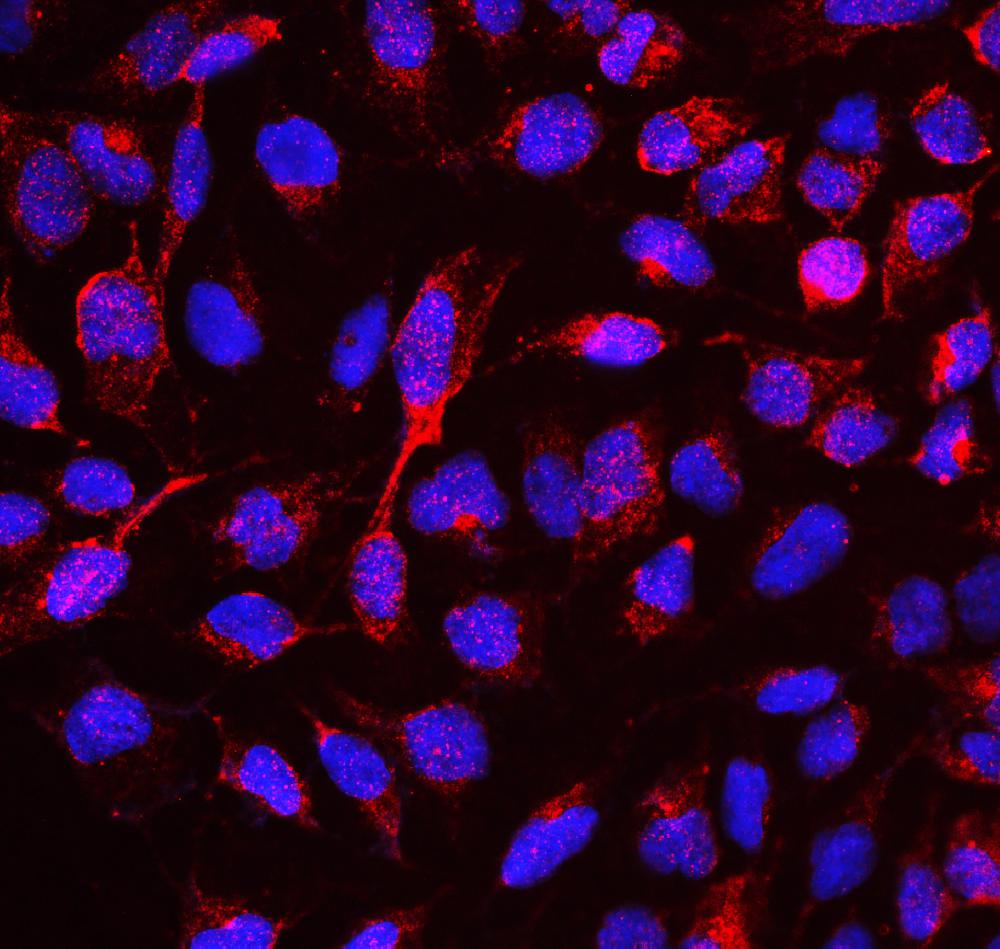
ICC/IF analysis of RBMS3 using anti- RBMS3 antibody. RBMS3 was detected in immunocytochemical section of U2OS cell. Enzyme antigen retrieval was performed using IHC enzyme antigen retrieval reagent rabbit anti- RBMS3 Antibody . Cy3 Conjugated Goat Anti-Rabbit IgG was used as secondary antibody The section was counterstained with DAPI. Visualize using a fluorescence microscope and filter sets appropriate for the label used.| Product Name | Rabbit polyclonal antibody to RBMS3 |
|---|---|
| Antibody Type | Primary Antibodies |
| Immunogen | E. coli-derived human RBMS3 recombinant protein (Position: T350-P437). Human RBMS3 shares 92% amino acid (aa) sequence identity with both mouse and rat RBMS3. |
| Clonality | Polyclonal |
|---|---|
| Isotype | IgG |
| Host Species | Rabbit |
| Tested Applications | ICC/IFIHCWB |
| WB:1:500-1:2000 IHC:1:50-1:400 ICC/IF:1:50-1:400 |
|
| Species Reactivity | HumanMouseRat |
| Concentration | 0.5mg/ml |
| Purification | Affinity purified |
| Gene Symbol | RBMS3 |
|---|---|
| Gene Full Name | RNA binding motif single stranded interacting protein 3 |
| Gene Summary | This gene encodes an RNA-binding protein that belongs to the c-myc gene single-strand binding protein family. These proteins are characterized by the presence of two sets of ribonucleoprotein consensus sequence (RNP-CS) that contain conserved motifs, RNP1 and RNP2, originally described in RNA binding proteins, and required for DNA binding. These proteins have been implicated in such diverse functions as DNA replication, gene transcription, cell cycle progression and apoptosis. The encoded protein was isolated by virtue of its binding to an upstream element of the alpha2(I) collagen promoter. The observation that this protein localizes mostly in the cytoplasm suggests that it may be involved in a cytoplasmic function such as controlling RNA metabolism, rather than transcription. Multiple alternatively spliced transcript variants encoding different isoforms have been found for this gene. [provided by RefSeq, Apr 2010] |
| Alternative Names | RNA binding motif single stranded interacting protein 3 RBMS3 |
| Molecular Weight(MW) | 48 kDa(Observed band size: 55 kDa) |
| Cellular Localization | Cytoplasm |
WB
Western blot analysis of RBMS3 using anti-RBMS3 antibody. The sample well of each lane was loaded with 30 ug of sample under reducing conditions. Lane 1: human U2OS whole cell lysates, Lane 2: human PC-3 whole cell lysates, Lane 3: human U-87MG whole cell lysates, Lane 4: human A549 whole cell lysates, Lane 5: human Hela whole cell lysates. After electrophoresis, proteins were transferred to a membrane. Then the membrane was incubated with rabbit anti-RBMS3 antigen affinity purified polyclonal antibod at a dilution of 1:1000 and probed with a goat anti-rabbit IgG-HRP secondary antibody. The signal is developed using ECL Plus Western Blotting Substrate.
IHC
IHC analysis of RBMS3 using anti-RBMS3 antibody. RBMS3 was detected in a paraffin-embedded section of human oesophagus squama cancer tissue. Biotinylated goat anti-rabbit IgG was used as secondary antibody. The tissue section was incubated with rabbit anti-RBMS3 Antibody at a dilution of 1:200 and developed using Strepavidin-Biotin-Complex (SABC) with DAB as the chromogen.
IHC
IHC analysis of RBMS3 using anti-RBMS3 antibody. RBMS3 was detected in a paraffin-embedded section of mouse brain tissue. Biotinylated goat anti-rabbit IgG was used as secondary antibody. The tissue section was incubated with rabbit anti-RBMS3 Antibody at a dilution of 1:200 and developed using Strepavidin-Biotin-Complex (SABC) with DAB as the chromogen.
IHC
IHC analysis of RBMS3 using anti-RBMS3 antibody. RBMS3 was detected in a paraffin-embedded section of rat brain tissue. Biotinylated goat anti-rabbit IgG was used as secondary antibody. The tissue section was incubated with rabbit anti-RBMS3 Antibody at a dilution of 1:200 and developed using Strepavidin-Biotin-Complex (SABC) with DAB as the chromogen.
ICC/IF
ICC/IF analysis of RBMS3 using anti- RBMS3 antibody. RBMS3 was detected in immunocytochemical section of U2OS cell. Enzyme antigen retrieval was performed using IHC enzyme antigen retrieval reagent rabbit anti- RBMS3 Antibody . Cy3 Conjugated Goat Anti-Rabbit IgG was used as secondary antibody The section was counterstained with DAPI. Visualize using a fluorescence microscope and filter sets appropriate for the label used.| Application Notes | WB:1:500-1:2000 IHC:1:50-1:400 ICC/IF:1:50-1:400 |
|---|
| Form | Liquid |
|---|---|
| Storage Instructions | 12 months from date of receipt, -20℃ as supplied. 6 months 2 to 8℃ after reconstitution. Avoid repeated freezing and thawing. |
| Storage Buffer | 500ug/ml antibody with PBS, 0.02% NaN3, 1 mg/ml BSA and 50% glycerol. |


