WB
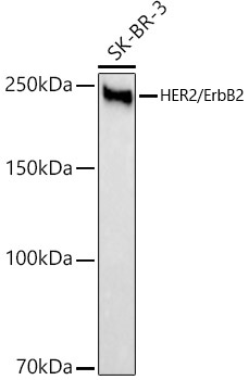
Western blot analysis of lysates from SK-BR-3 cells, using HER2/ErbB2 Rabbit mAb at1:2000 dilution. Secondary antibody: HRP-conjugated Goat anti-Rabbit IgG (H+L) at 1:10000 dilution. Lysates/proteins: 25μg per lane. Blocking buffer: 3% nonfat dry milk in TBST. Detection: ECL Basic Kit. Exposure time: 1s.IHC
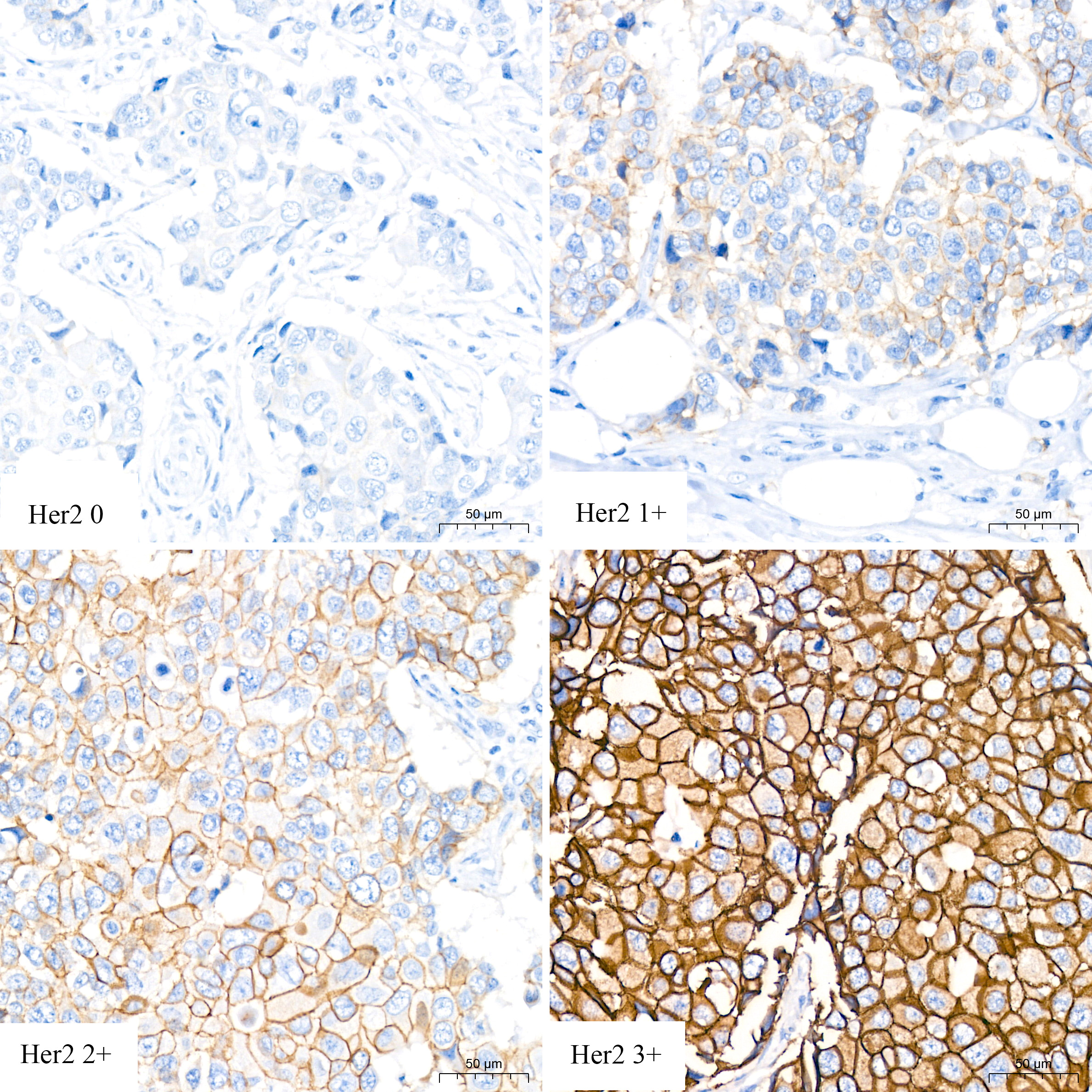
Immunohistochemistry analysis of paraffin embedded Human breast cancer using HER2/ErbB2 Rabbit mAb at dilution of 1:200 (40x lens). High pressure antigen retrieval performed with 0.01M Citrate buffer (pH 6.0) prior to IHC staining.IHC
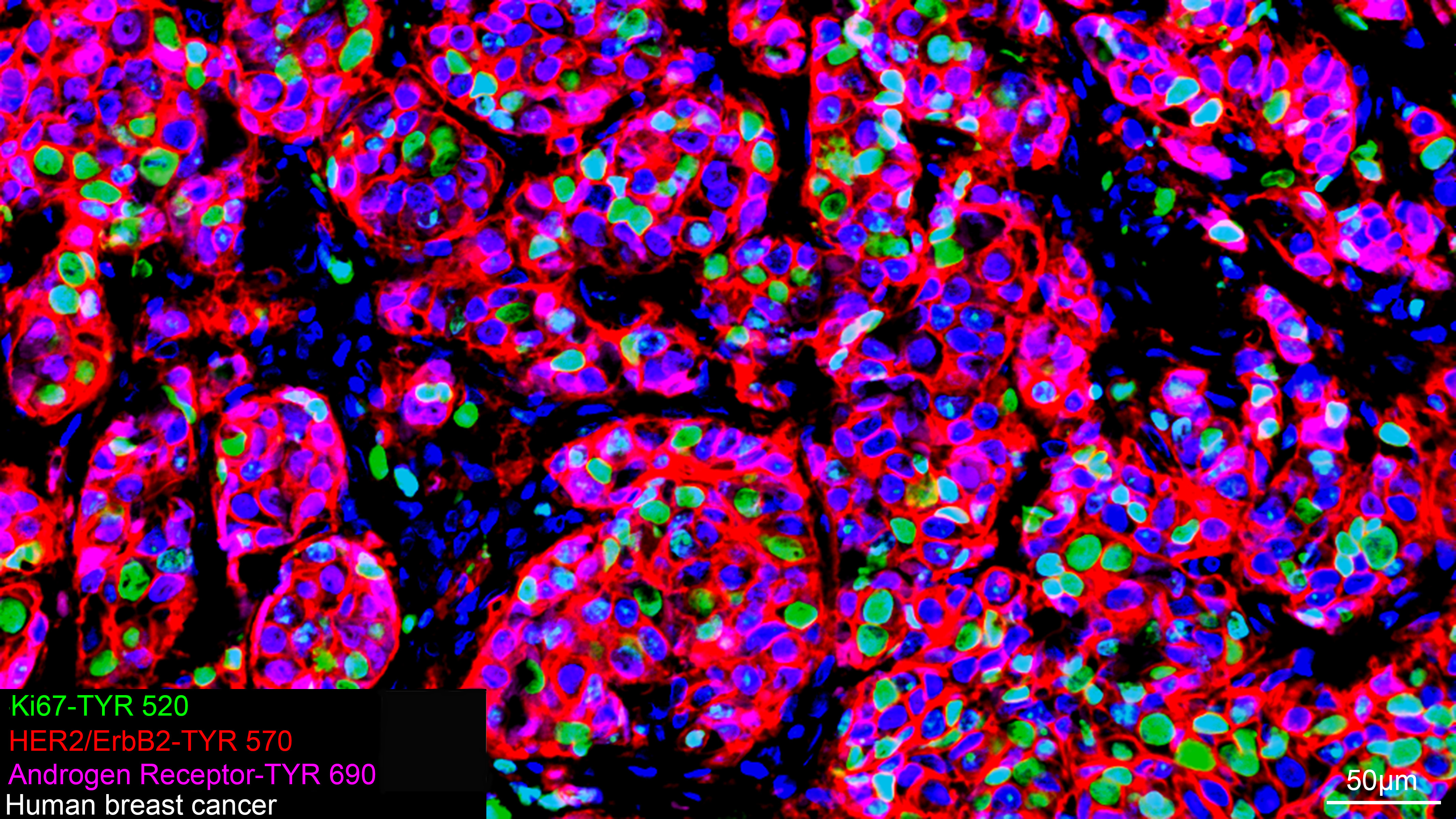
The multiplex IHC analysis on paraffin embedded Human breast cancer tissue using the following specific primary antibodies and tyramide signal amplification (TSA) reagents : Ki67 Rabbit mAb (1:500) with TSA-TYR-520 (Green), HER2/ErbB2 Rabbit mAb (1:200) with TSA-TYR-570 (Red), and Androgen Receptor Rabbit mAb (1:400) with TSA-TYR-690 (Magenta). DAPI (Blue) was used for nuclear staining. Prior to multiplex IHC staining, high-pressure antigen retrieval was performed using 0.01M citrate buffer at pH 6.0. The analysis was completed using a 20x objective lens.IF-P
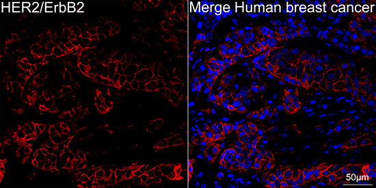
Confocal imaging of paraffin-embedded Human breast cancer tissue using HER2/ErbB2 Rabbit mAb (dilution 1:200) followed by a further incubation with Cy3 Goat Anti-Rabbit IgG (H+L) (dilution 1:500) (Red). DAPI was used for nuclear staining (Blue). High pressure antigen retrieval performed with 0.01M Citrate Buffer (pH 6.0) prior to IF staining. Objective: 40x.| Product Name | HER2/ErbB2 Rabbit mAb |
|---|---|
| Antibody Type | Primary Antibodies |
| Immunogen | Recombinant protein (or fragment).This information is considered to be commercially sensitive. |
| Clonality | Monoclonal |
|---|---|
| Isotype | IgG |
| Host Species | Rabbit |
| Tested Applications | IF-PIHCWB |
| WB:1:2000 - 1:4000 IHC:1:200 - 1:800 IF-P:1:200 - 1:800 |
|
| Species Reactivity | Human |
| Concentration | 1mg/ml |
| Purification | Affinity purified |
| Gene Symbol | ERBB2 |
|---|---|
| Gene Synonyms | NEU NGL HER2 TKR1 CD340 HER-2 VSCN2 MLN 19 MLN-19 c-ERB2 c-ERB-2 HER-2/neu p185(erbB2) |
| Gene Full Name | erb-b2 receptor tyrosine kinase 2 |
| Gene Summary | This gene encodes a member of the epidermal growth factor (EGF) receptor family of receptor tyrosine kinases. This protein has no ligand binding domain of its own and therefore cannot bind growth factors. However, it does bind tightly to other ligand-bound EGF receptor family members to form a heterodimer, stabilizing ligand binding and enhancing kinase-mediated activation of downstream signalling pathways, such as those involving mitogen-activated protein kinase and phosphatidylinositol-3 kinase. Allelic variations at amino acid positions 654 and 655 of isoform a (positions 624 and 625 of isoform b) have been reported, with the most common allele, Ile654/Ile655, shown here. Amplification and/or overexpression of this gene has been reported in numerous cancers, including breast and ovarian tumors. Alternative splicing results in several additional transcript variants, some encoding different isoforms and others that have not been fully characterized. [provided by RefSeq, Jul 2008] |
| Alternative Names | NEU NGL HER2 TKR1 CD340 HER-2 VSCN2 MLN 19 MLN-19 c-ERB2 c-ERB-2 HER-2/neu p185(erbB2) HER2/ErbB2 |
| Molecular Weight(MW) | 138 kDa(Observed band size:185 kDa) |
| Cellular Localization | Cell membrane, Cytoplasm, Single-pass type I membrane protein. |
WB
Western blot analysis of lysates from SK-BR-3 cells, using HER2/ErbB2 Rabbit mAb at1:2000 dilution. Secondary antibody: HRP-conjugated Goat anti-Rabbit IgG (H+L) at 1:10000 dilution. Lysates/proteins: 25μg per lane. Blocking buffer: 3% nonfat dry milk in TBST. Detection: ECL Basic Kit. Exposure time: 1s.
IHC
Immunohistochemistry analysis of paraffin embedded Human breast cancer using HER2/ErbB2 Rabbit mAb at dilution of 1:200 (40x lens). High pressure antigen retrieval performed with 0.01M Citrate buffer (pH 6.0) prior to IHC staining.
IHC
The multiplex IHC analysis on paraffin embedded Human breast cancer tissue using the following specific primary antibodies and tyramide signal amplification (TSA) reagents : Ki67 Rabbit mAb (1:500) with TSA-TYR-520 (Green), HER2/ErbB2 Rabbit mAb (1:200) with TSA-TYR-570 (Red), and Androgen Receptor Rabbit mAb (1:400) with TSA-TYR-690 (Magenta). DAPI (Blue) was used for nuclear staining. Prior to multiplex IHC staining, high-pressure antigen retrieval was performed using 0.01M citrate buffer at pH 6.0. The analysis was completed using a 20x objective lens.
IF-P
Confocal imaging of paraffin-embedded Human breast cancer tissue using HER2/ErbB2 Rabbit mAb (dilution 1:200) followed by a further incubation with Cy3 Goat Anti-Rabbit IgG (H+L) (dilution 1:500) (Red). DAPI was used for nuclear staining (Blue). High pressure antigen retrieval performed with 0.01M Citrate Buffer (pH 6.0) prior to IF staining. Objective: 40x.| Application Notes | WB:1:2000 - 1:4000 IHC:1:200 - 1:800 IF-P:1:200 - 1:800 |
|---|
| Form | Liquid |
|---|---|
| Storage Instructions | Store at -20℃. Avoid freeze / thaw cycles. |
| Storage Buffer | PBS containing 50% glycerol and 0.05% BSA, preserved with proclin300 or sodium azide (as specified on the Certificate of Analysis), pH 7.3. |


