WB
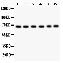
Western blot analysis of PKC Iota/PRKCI using anti-PKC Iota/PRKCI antibody. The sample well of each lane was loaded with 30 ug of sample under reducing conditions. After electrophoresis, proteins were transferred to a membrane. Then the membrane was incubated with rabbit anti-PKC Iota/PRKCI antigen affinity purified polyclonal antibody at a dilution of 1:1000 and probed with a goat anti-rabbit IgG-HRP secondary antibody. The signal is developed using ECL Plus Western Blotting Substrate.IHC
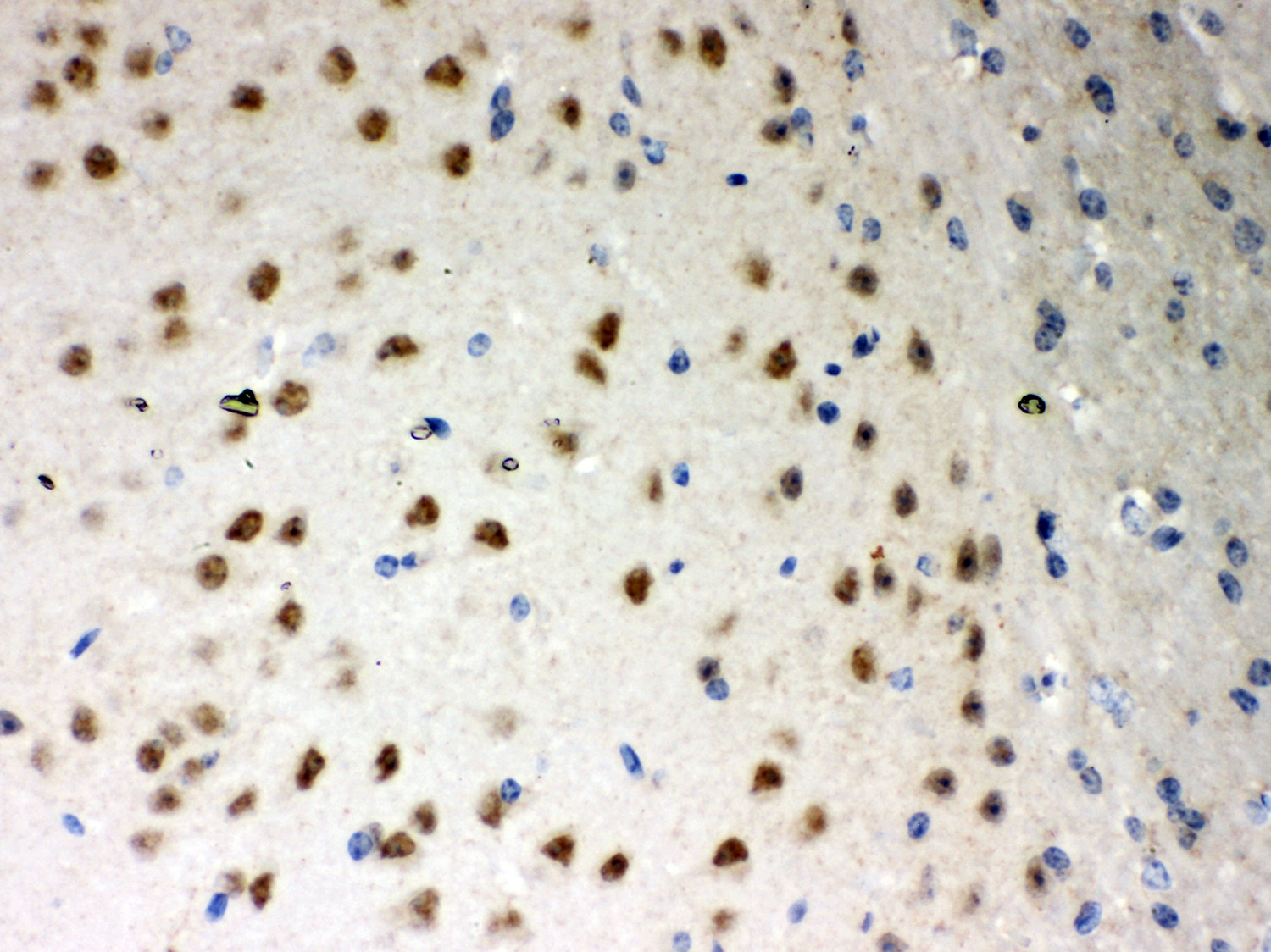
IHC analysis of PKC Iota/PRKCI using anti-PKC Iota/PRKCI antibody. PKC Iota/PRKCI was detected in a paraffin-embedded section of mouse brain tissue. Biotinylated goat anti-rabbit IgG was used as secondary antibody. The tissue section was incubated with rabbit anti-PKC Iota/PRKCI Antibody at a dilution of 1:200 and developed using Strepavidin-Biotin-Complex (SABC) with DAB as the chromogen.IHC
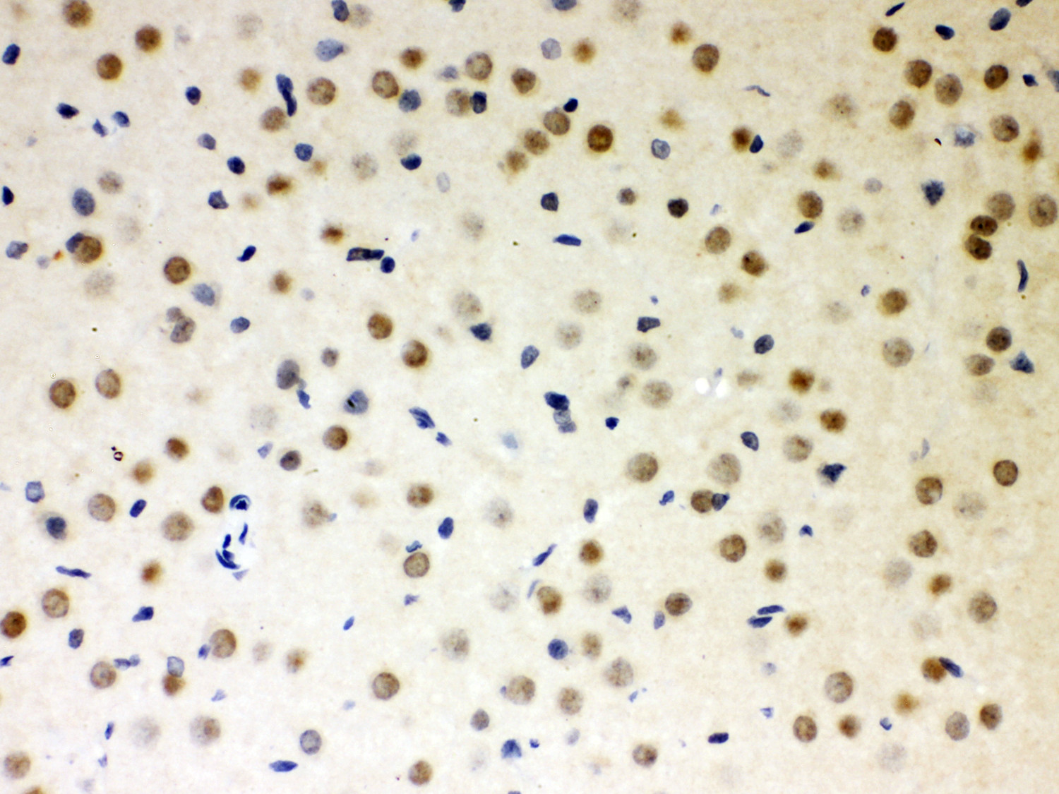
IHC analysis of PKC Iota/PRKCI using anti-PKC Iota/PRKCI antibody. PKC Iota/PRKCI was detected in a paraffin-embedded section of rat brain tissue. Biotinylated goat anti-rabbit IgG was used as secondary antibody. The tissue section was incubated with rabbit anti-PKC Iota/PRKCI Antibody at a dilution of 1:200 and developed using Strepavidin-Biotin-Complex (SABC) with DAB as the chromogen.IHC
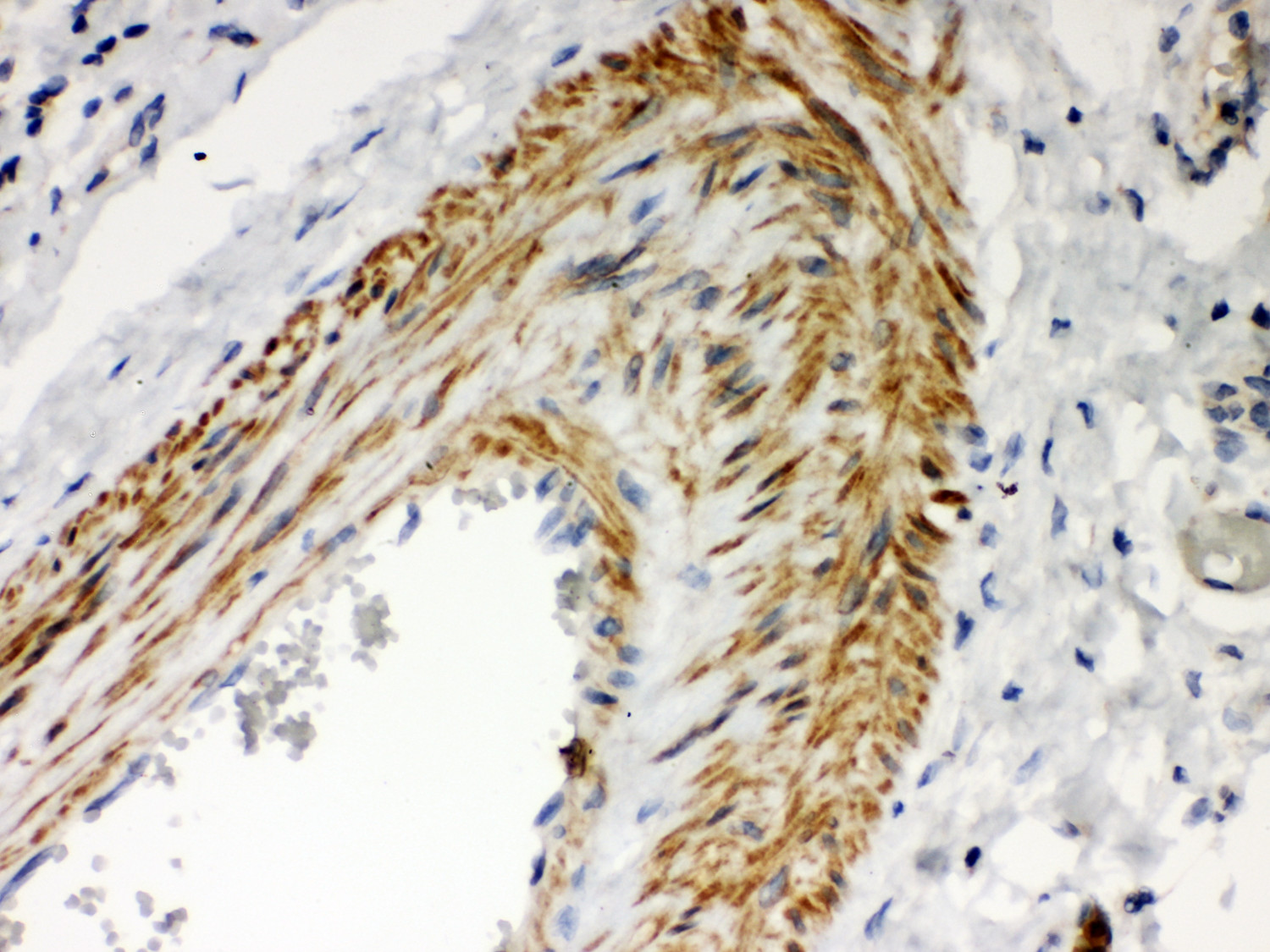
IHC analysis of PKC Iota/PRKCI using anti-PKC Iota/PRKCI antibody. PKC Iota/PRKCI was detected in a paraffin-embedded section of human lung cancer tissue. Biotinylated goat anti-rabbit IgG was used as secondary antibody. The tissue section was incubated with rabbit anti-PKC Iota/PRKCI Antibody at a dilution of 1:200 and developed using Strepavidin-Biotin-Complex (SABC) with DAB as the chromogen.| Product Name | Rabbit polyclonal antibody to PKC Iota/PRKCI |
|---|---|
| Antibody Type | Primary Antibodies |
| Immunogen | E.coli-derived human PKC iota recombinant protein (Position: D21-Q214). Human PKC iota shares 96% and 97% amino acid (aa) sequence identity with mouse and rat PKC iota, respectively. |
| Clonality | Polyclonal |
|---|---|
| Isotype | IgG |
| Host Species | Rabbit |
| Tested Applications | IHCWB |
| WB:1:500-1:2000 IHC:1:50-1:400 |
|
| Species Reactivity | HumanMouseRat |
| Concentration | 0.5mg/ml |
| Purification | Affinity purified |
| Gene Symbol | PRKCI |
|---|---|
| Gene Synonyms | PKCI DXS1179E nPKC-iota |
| Gene Full Name | protein kinase C iota |
| Gene Summary | This gene encodes a member of the protein kinase C (PKC) family of serine/threonine protein kinases. The PKC family comprises at least eight members, which are differentially expressed and are involved in a wide variety of cellular processes. This protein kinase is calcium-independent and phospholipid-dependent. It is not activated by phorbolesters or diacylglycerol. This kinase can be recruited to vesicle tubular clusters (VTCs) by direct interaction with the small GTPase RAB2, where this kinase phosphorylates glyceraldehyde-3-phosphate dehydrogenase (GAPD/GAPDH) and plays a role in microtubule dynamics in the early secretory pathway. This kinase is found to be necessary for BCL-ABL-mediated resistance to drug-induced apoptosis and therefore protects leukemia cells against drug-induced apoptosis. There is a single exon pseudogene mapped on chromosome X. [provided by RefSeq, Jul 2008] |
| Alternative Names | aPKC lambda/iota DXS1179E PKC iota PKCI PRKC lambda/iota PRKCI Protein kinase C iota type protein kinase C iota |
| Molecular Weight(MW) | 68 kDa |
| Cellular Localization | Cytoplasm,Endosome,Membrane,Nucleus. |
WB
Western blot analysis of PKC Iota/PRKCI using anti-PKC Iota/PRKCI antibody. The sample well of each lane was loaded with 30 ug of sample under reducing conditions. After electrophoresis, proteins were transferred to a membrane. Then the membrane was incubated with rabbit anti-PKC Iota/PRKCI antigen affinity purified polyclonal antibody at a dilution of 1:1000 and probed with a goat anti-rabbit IgG-HRP secondary antibody. The signal is developed using ECL Plus Western Blotting Substrate.
IHC
IHC analysis of PKC Iota/PRKCI using anti-PKC Iota/PRKCI antibody. PKC Iota/PRKCI was detected in a paraffin-embedded section of mouse brain tissue. Biotinylated goat anti-rabbit IgG was used as secondary antibody. The tissue section was incubated with rabbit anti-PKC Iota/PRKCI Antibody at a dilution of 1:200 and developed using Strepavidin-Biotin-Complex (SABC) with DAB as the chromogen.
IHC
IHC analysis of PKC Iota/PRKCI using anti-PKC Iota/PRKCI antibody. PKC Iota/PRKCI was detected in a paraffin-embedded section of rat brain tissue. Biotinylated goat anti-rabbit IgG was used as secondary antibody. The tissue section was incubated with rabbit anti-PKC Iota/PRKCI Antibody at a dilution of 1:200 and developed using Strepavidin-Biotin-Complex (SABC) with DAB as the chromogen.
IHC
IHC analysis of PKC Iota/PRKCI using anti-PKC Iota/PRKCI antibody. PKC Iota/PRKCI was detected in a paraffin-embedded section of human lung cancer tissue. Biotinylated goat anti-rabbit IgG was used as secondary antibody. The tissue section was incubated with rabbit anti-PKC Iota/PRKCI Antibody at a dilution of 1:200 and developed using Strepavidin-Biotin-Complex (SABC) with DAB as the chromogen.| Application Notes | WB:1:500-1:2000 IHC:1:50-1:400 |
|---|
| Form | Liquid |
|---|---|
| Storage Instructions | 12 months from date of receipt,-20℃ as supplied. |
| Storage Buffer | 500 ug/ml antibody with PBS, 0.02% NaN3, 1 mg/ml BSA and 50% glycerol. |


