WB
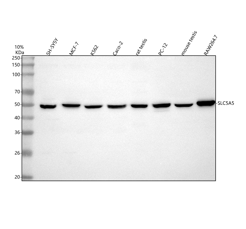
Western blot analysis of SLC5A5 using anti-SLC5A5 antibody. The sample well of each lane was loaded with 30 ug of sample under reducing conditions. Lane 1: human SH-SY5Y whole cell lysates, Lane 2: human MCF-7 whole cell lysates, Lane 3: human K562 whole cell lysates, Lane 4: human Caco-2 whole cell lysates, Lane 5: rat testis tissue lysates, Lane 6: rat PC-12 whole cell lysates, Lane 7: mouse testis tissue lysates, Lane 8: mouse RAW264.7 whole cell lysates. After electrophoresis, proteins were transferred to a membrane. Then the membrane was incubated with rabbit anti-SLC5A5 antigen affinity purified polyclonal antibody at a dilution of 1:1000 and probed with a goat anti-rabbit IgG-HRP secondary antibody. The signal is developed using ECL Plus Western Blotting Substrate.IHC
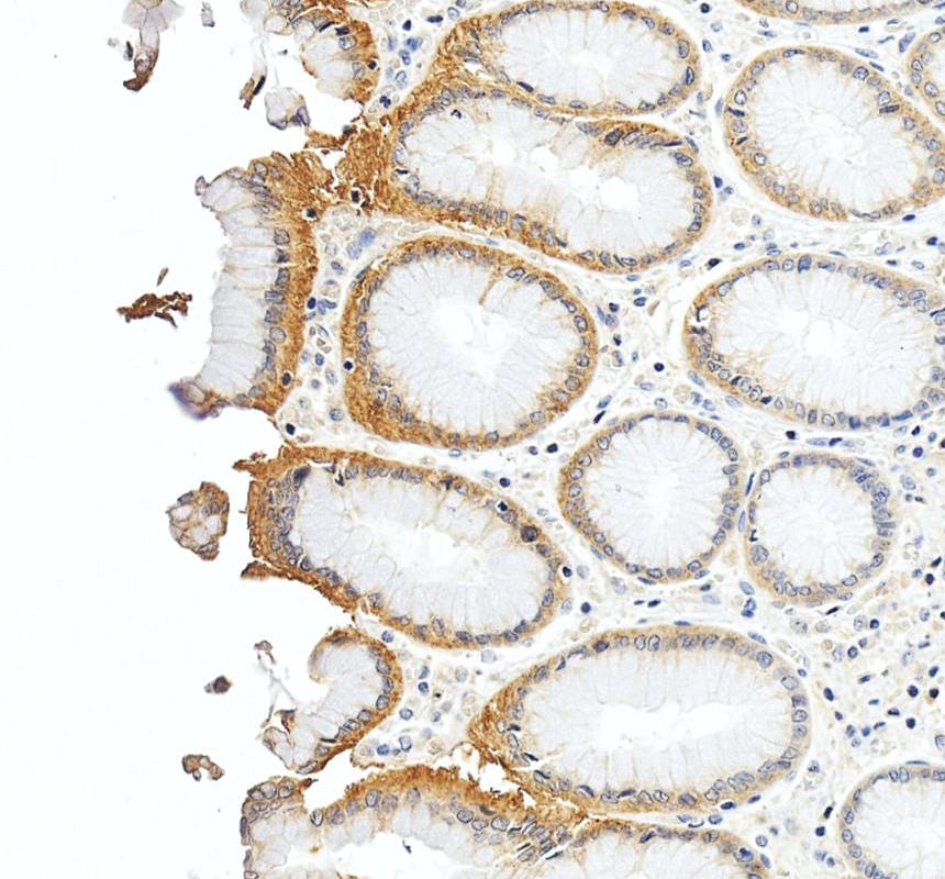
SLC5A5 was detected in a paraffin-embedded section of human stomach tissue. The tissue section was incubated with rabbit anti-SLC5A5 Antibody at a dilution of 1:200 and developed using HRP Conjugated Rabbit IgG Super Vision Assay Kit with DAB as the chromogen.IHC
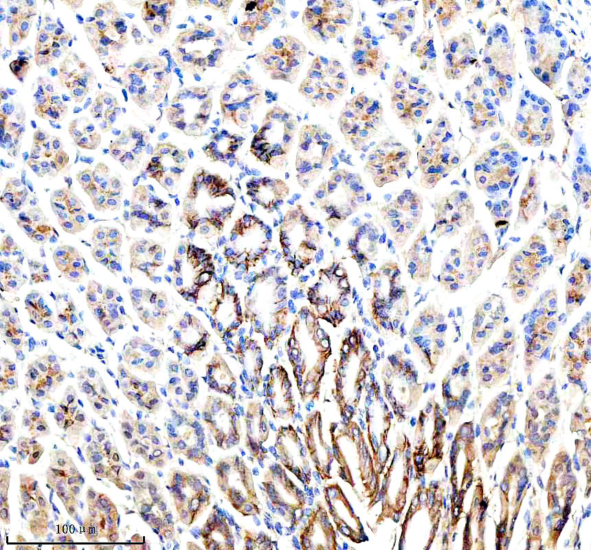
SLC5A5 was detected in a paraffin-embedded section of mouse stomach tissue. The tissue section was incubated with rabbit anti-SLC5A5 Antibody at a dilution of 1:200 and developed using HRP Conjugated Rabbit IgG Super Vision Assay Kit with DAB as the chromogen.IHC
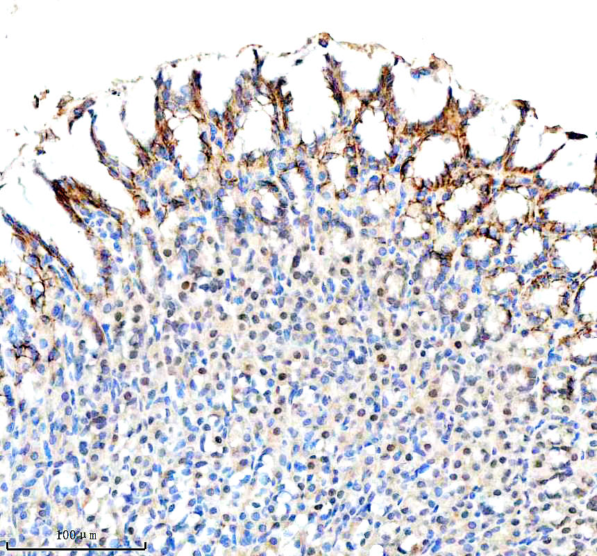
SLC5A5 was detected in a paraffin-embedded section of rat stomach tissue. The tissue section was incubated with rabbit anti-SLC5A5 Antibody at a dilution of 1:200 and developed using HRP Conjugated Rabbit IgG Super Vision Assay Kit with DAB as the chromogen.| Product Name | Rabbit polyclonal antibody to SLC5A5 |
|---|---|
| Antibody Type | Primary Antibodies |
| Immunogen | E.coli-derived human SLC5A5 recombinant protein (Position: L541-L643). |
| Clonality | Polyclonal |
|---|---|
| Isotype | IgG |
| Host Species | Rabbit |
| Tested Applications | IHCWB |
| WB:1:500-1:2000 IHC:1:50-1:400 |
|
| Species Reactivity | HumanMouseRat |
| Concentration | 0.5mg/ml |
| Purification | Affinity purified |
| Gene Symbol | SLC5A5 |
|---|---|
| Gene Synonyms | NIS TDH1 |
| Gene Full Name | solute carrier family 5 member 5 |
| Gene Summary | This gene encodes a member of the sodium glucose cotransporter family. The encoded protein is responsible for the uptake of iodine in tissues such as the thyroid and lactating breast tissue. The iodine taken up by the thyroid is incorporated into the metabolic regulators triiodothyronine (T3) and tetraiodothyronine (T4). Mutations in this gene are associated with thyroid dyshormonogenesis 1.[provided by RefSeq, Sep 2009] |
| Alternative Names | Sodium/iodide cotransporter |
| Molecular Weight(MW) | 50 kDa |
| Cellular Localization | Cell membrane,Cytoplasm. |
WB
Western blot analysis of SLC5A5 using anti-SLC5A5 antibody. The sample well of each lane was loaded with 30 ug of sample under reducing conditions. Lane 1: human SH-SY5Y whole cell lysates, Lane 2: human MCF-7 whole cell lysates, Lane 3: human K562 whole cell lysates, Lane 4: human Caco-2 whole cell lysates, Lane 5: rat testis tissue lysates, Lane 6: rat PC-12 whole cell lysates, Lane 7: mouse testis tissue lysates, Lane 8: mouse RAW264.7 whole cell lysates. After electrophoresis, proteins were transferred to a membrane. Then the membrane was incubated with rabbit anti-SLC5A5 antigen affinity purified polyclonal antibody at a dilution of 1:1000 and probed with a goat anti-rabbit IgG-HRP secondary antibody. The signal is developed using ECL Plus Western Blotting Substrate.
IHC
SLC5A5 was detected in a paraffin-embedded section of human stomach tissue. The tissue section was incubated with rabbit anti-SLC5A5 Antibody at a dilution of 1:200 and developed using HRP Conjugated Rabbit IgG Super Vision Assay Kit with DAB as the chromogen.
IHC
SLC5A5 was detected in a paraffin-embedded section of mouse stomach tissue. The tissue section was incubated with rabbit anti-SLC5A5 Antibody at a dilution of 1:200 and developed using HRP Conjugated Rabbit IgG Super Vision Assay Kit with DAB as the chromogen.
IHC
SLC5A5 was detected in a paraffin-embedded section of rat stomach tissue. The tissue section was incubated with rabbit anti-SLC5A5 Antibody at a dilution of 1:200 and developed using HRP Conjugated Rabbit IgG Super Vision Assay Kit with DAB as the chromogen.| Application Notes | WB:1:500-1:2000 IHC:1:50-1:400 |
|---|
| Form | Liquid |
|---|---|
| Storage Instructions | 12 months from date of receipt,-20℃ as supplied. |
| Storage Buffer | 500 ug/ml antibody with PBS, 0.02% NaN3, 1 mg/ml BSA and 50% glycerol. |


