WB
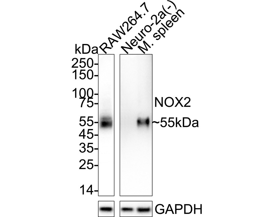
Relative expression (RE) Western blot analysis of NOX2 / gp91phox on different lysates with Rabbit anti-NOX2 / gp91phox antibody at 1/5,000 dilution. Exposure time: Lane 1: 16 seconds; Lane 2-3: 46 seconds; 4-20% SDS-PAGE gel. Proteins were transferred to a PVDF membrane and blocked with 5% NFDM/TBST for 1 hour at room temperature. The primary antibody at 1/5,000 dilution was used in 5% NFDM/TBST at 4℃ overnight. Goat Anti-Rabbit IgG - HRP Secondary Antibody at 1/50,000 dilution was used for 1 hour at room temperature.IHC
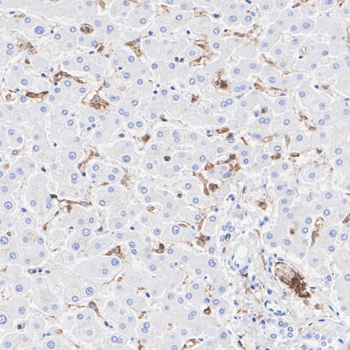
Immunohistochemical analysis of paraffin-embedded human liver tissue with Rabbit anti-NOX2 / gp91phox antibody at 1/1,000 dilution. The section was pre-treated using heat mediated antigen retrieval with Tris-EDTA buffer (pH 9.0) for 20 minutes. The tissues were blocked in 1% BSA for 20 minutes at room temperature, washed with ddH2O and PBS, and then probed with the primary antibody at 1/1,000 dilution for 1 hour at room temperature. The detection was performed using an HRP conjugated compact polymer system. DAB was used as the chromogen. Tissues were counterstained with hematoxylin and mounted with DPX.IHC
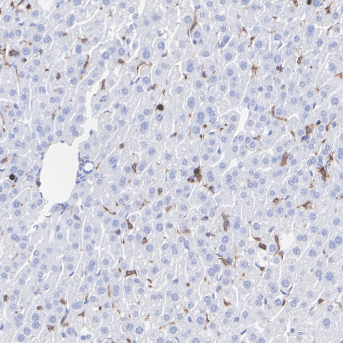
Immunohistochemical analysis of paraffin-embedded mouse liver tissue with Rabbit anti-NOX2 / gp91phox antibody at 1/1,000 dilution. The section was pre-treated using heat mediated antigen retrieval with Tris-EDTA buffer (pH 9.0) for 20 minutes. The tissues were blocked in 1% BSA for 20 minutes at room temperature, washed with ddH2O and PBS, and then probed with the primary antibody at 1/1,000 dilution for 1 hour at room temperature. The detection was performed using an HRP conjugated compact polymer system. DAB was used as the chromogen. Tissues were counterstained with hematoxylin and mounted with DPX.IHC
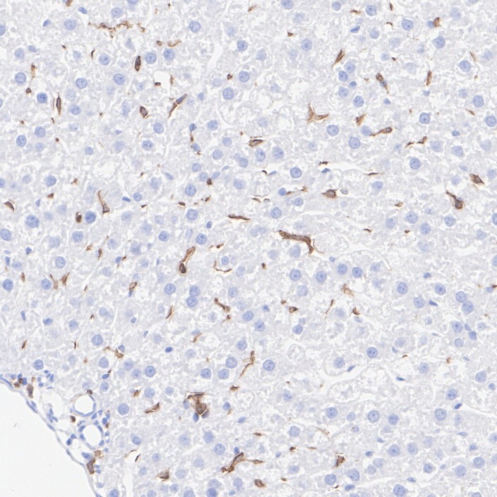
Immunohistochemical analysis of paraffin-embedded rat liver tissue with Rabbit anti-NOX2 / gp91phox antibody at 1/1,000 dilution. The section was pre-treated using heat mediated antigen retrieval with Tris-EDTA buffer (pH 9.0) for 20 minutes. The tissues were blocked in 1% BSA for 20 minutes at room temperature, washed with ddH2O and PBS, and then probed with the primary antibody at 1/1,000 dilution for 1 hour at room temperature. The detection was performed using an HRP conjugated compact polymer system. DAB was used as the chromogen. Tissues were counterstained with hematoxylin and mounted with DPX.ICC/IF
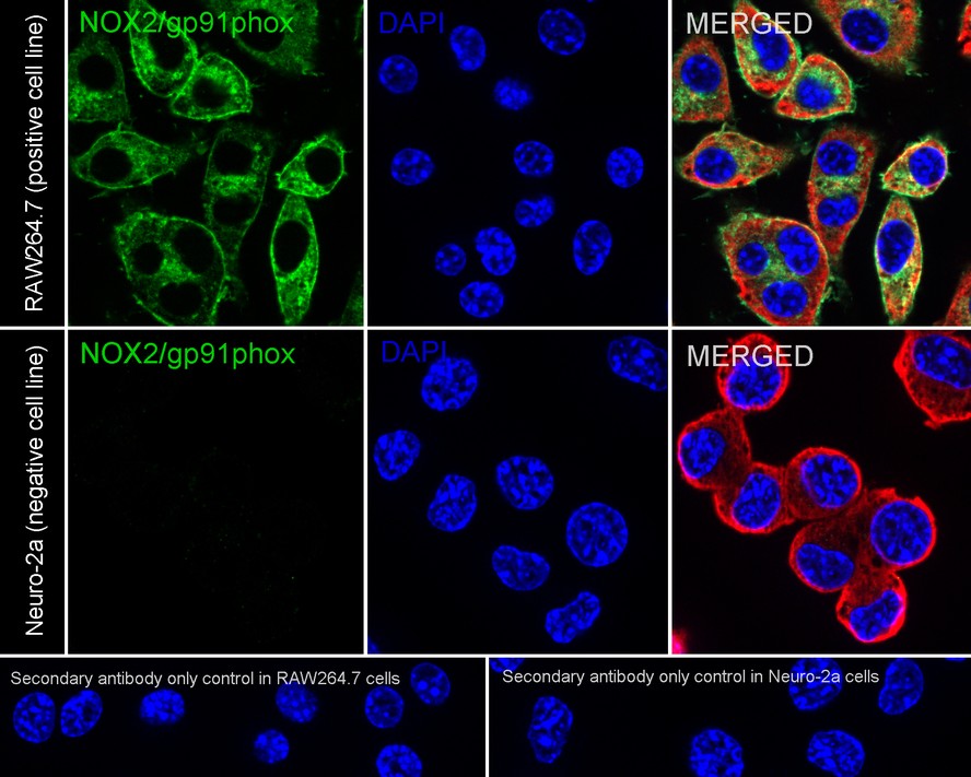
Relative expression (RE) Immunocytochemistry analysis of RAW264.7 (positive) and Neuro-2a (negative) labeling NOX2 / gp91phox with Rabbit anti-NOX2 / gp91phox antibody at 1/500 dilution. Cells were fixed in 4% paraformaldehyde for 15 minutes at room temperature, permeabilized with 0.1% Triton X-100 in PBS for 15 minutes at room temperature, then blocked with 1% BSA in 10% negative goat serum for 1 hour at room temperature. Cells were then incubated with Rabbit anti-NOX2 / gp91phox antibody at 1/500 dilution in 1% BSA in PBST overnight at 4 ℃. Goat Anti-Rabbit IgG H&L (488) was used as the secondary antibody at 1/1,000 dilution. PBS instead of the primary antibody was used as the secondary antibody only control. Nuclear DNA was labelled in blue with DAPI. Beta tubulin (red) was stained at 1/100 dilution overnight at +4℃. Goat Anti-Mouse IgG H&L (594) was used as the secondary antibody at 1/1,000 dilution.IF-F
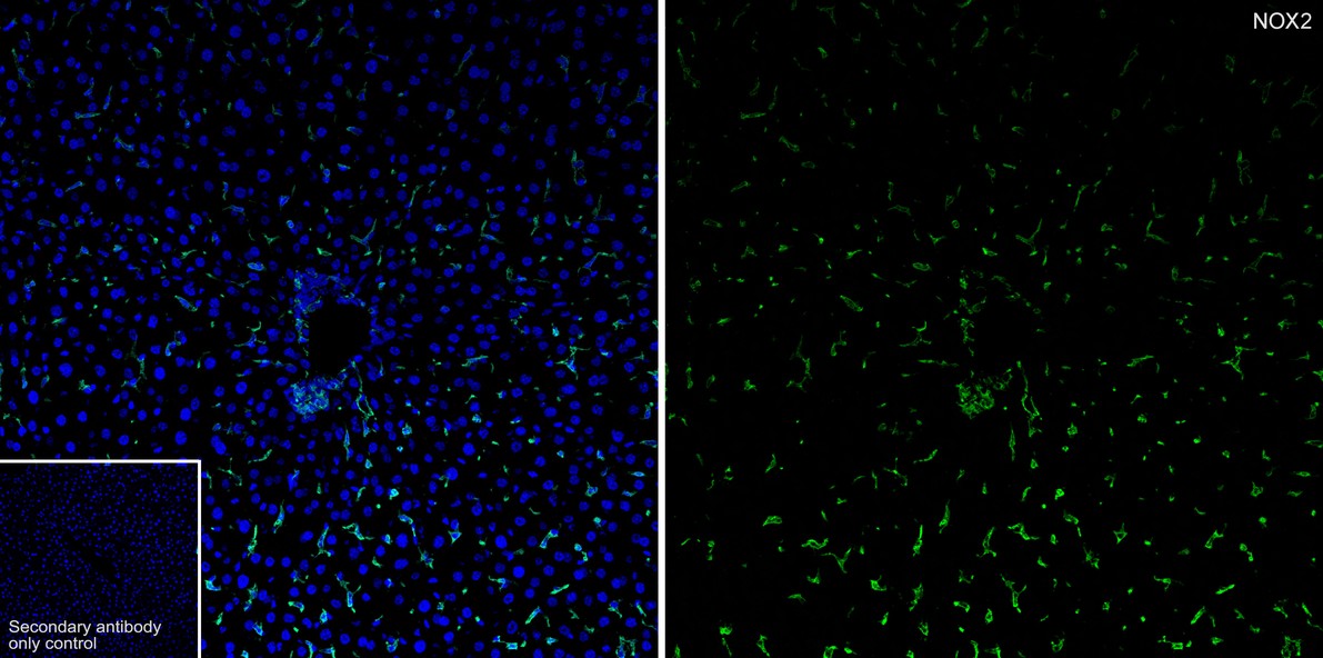
Immunofluorescence analysis of frozen mouse liver tissue with Rabbit anti-NOX2 / gp91phox antibody at 1/500 dilution. The section was pre-treated using heat mediated antigen retrieval with sodium citrate buffer (pH 6.0) for about 2 minutes in microwave oven. The tissues were blocked in 10% negative goat serum for 1 hour at room temperature, washed with PBS, and then probed with the primary antibody (green) at 1/500 dilution overnight at 4 ℃, washed with PBS. Goat Anti-Rabbit IgG H&L (488) was used as the secondary antibody at 1/1,000 dilution. Nuclei were counterstained with DAPI (blue).IP
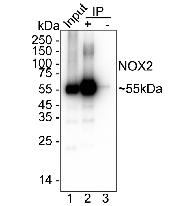
NOX2 / gp91phox was immunoprecipitated from 0.2 mg RAW264.7 cell lysate with NOX2 / gp91phox Recombinant Rabbit Monoclonal Antibody at 2 µg/10 µl beads. Western blot was performed from the immunoprecipitate using NOX2 / gp91phox Recombinant Rabbit Monoclonal Antibody at 1/2,000 dilution. HRP Conjugated Anti-Rabbit IgG for IP Nano-secondary antibody at 1/5,000 dilution was used for 1 hour at room temperature. Blocking/Dilution buffer: 5% NFDM/TBST Exposure time: 1 3 seconds;| Product Name | NOX2 / gp91phox Recombinant Rabbit Monoclonal Antibody |
|---|---|
| Antibody Type | Primary Antibodies |
| Clonality | Monoclonal |
|---|---|
| Isotype | IgG |
| Host Species | Rabbit |
| Tested Applications | ICC/IFIF-FIHCIPWB |
| WB:1:5000 IHC:1:1000 ICC/IF:1:500 IF-F:1:500 IP:1-2μg/sample |
|
| Species Reactivity | HumanMouseRat |
| Concentration | 1mg/ml |
| Purification | Protein A |
| Gene Symbol | CYBB |
|---|---|
| Gene Synonyms | CGD CGDX NOX2 IMD34 AMCBX2 GP91-1 GP91PHOX p91-PHOX GP91-PHOX |
| Gene Full Name | cytochrome b-245 beta chain |
| Gene Summary | Cytochrome b (-245) is composed of cytochrome b alpha (CYBA) and beta (CYBB) chain. It has been proposed as a primary component of the microbicidal oxidase system of phagocytes. CYBB deficiency is one of five described biochemical defects associated with chronic granulomatous disease (CGD). In this disorder, there is decreased activity of phagocyte NADPH oxidase; neutrophils are able to phagocytize bacteria but cannot kill them in the phagocytic vacuoles. The cause of the killing defect is an inability to increase the cell's respiration and consequent failure to deliver activated oxygen into the phagocytic vacuole. [provided by RefSeq, Jul 2008] |
| Alternative Names | AMCBX2 antibody CGD antibody CGD91-phox antibody CY24B_HUMAN antibody CYBB antibody Cytochrome b 245 beta polypeptide antibody Cytochrome b(558) beta chain antibody Cytochrome b(558) subunit beta antibody Cytochrome b-245 heavy chain antibody Cytochrome b558 subunit beta antibody GP91 PHOX antibody gp91-1 antibody gp91-phox antibody GP91PHOX antibody Heme-binding membrane glycoprotein gp91phox antibody NADPH oxidase 2 antibody Neutrophil cytochrome b 91 kDa polypeptide antibody NOX2 antibody p22 phagocyte B-cytochrome antibody P91 PHOX antibody p91-PHOX antibody Superoxide-generating NADPH oxidase heavy chain subunit antibody |
| Molecular Weight(MW) | 65 kDa(Observed band size: 55 kDa) |
| Cellular Localization | Cell membrane. |
WB
Relative expression (RE) Western blot analysis of NOX2 / gp91phox on different lysates with Rabbit anti-NOX2 / gp91phox antibody at 1/5,000 dilution. Exposure time: Lane 1: 16 seconds; Lane 2-3: 46 seconds; 4-20% SDS-PAGE gel. Proteins were transferred to a PVDF membrane and blocked with 5% NFDM/TBST for 1 hour at room temperature. The primary antibody at 1/5,000 dilution was used in 5% NFDM/TBST at 4℃ overnight. Goat Anti-Rabbit IgG - HRP Secondary Antibody at 1/50,000 dilution was used for 1 hour at room temperature.
IHC
Immunohistochemical analysis of paraffin-embedded human liver tissue with Rabbit anti-NOX2 / gp91phox antibody at 1/1,000 dilution. The section was pre-treated using heat mediated antigen retrieval with Tris-EDTA buffer (pH 9.0) for 20 minutes. The tissues were blocked in 1% BSA for 20 minutes at room temperature, washed with ddH2O and PBS, and then probed with the primary antibody at 1/1,000 dilution for 1 hour at room temperature. The detection was performed using an HRP conjugated compact polymer system. DAB was used as the chromogen. Tissues were counterstained with hematoxylin and mounted with DPX.
IHC
Immunohistochemical analysis of paraffin-embedded mouse liver tissue with Rabbit anti-NOX2 / gp91phox antibody at 1/1,000 dilution. The section was pre-treated using heat mediated antigen retrieval with Tris-EDTA buffer (pH 9.0) for 20 minutes. The tissues were blocked in 1% BSA for 20 minutes at room temperature, washed with ddH2O and PBS, and then probed with the primary antibody at 1/1,000 dilution for 1 hour at room temperature. The detection was performed using an HRP conjugated compact polymer system. DAB was used as the chromogen. Tissues were counterstained with hematoxylin and mounted with DPX.
IHC
Immunohistochemical analysis of paraffin-embedded rat liver tissue with Rabbit anti-NOX2 / gp91phox antibody at 1/1,000 dilution. The section was pre-treated using heat mediated antigen retrieval with Tris-EDTA buffer (pH 9.0) for 20 minutes. The tissues were blocked in 1% BSA for 20 minutes at room temperature, washed with ddH2O and PBS, and then probed with the primary antibody at 1/1,000 dilution for 1 hour at room temperature. The detection was performed using an HRP conjugated compact polymer system. DAB was used as the chromogen. Tissues were counterstained with hematoxylin and mounted with DPX.
ICC/IF
Relative expression (RE) Immunocytochemistry analysis of RAW264.7 (positive) and Neuro-2a (negative) labeling NOX2 / gp91phox with Rabbit anti-NOX2 / gp91phox antibody at 1/500 dilution. Cells were fixed in 4% paraformaldehyde for 15 minutes at room temperature, permeabilized with 0.1% Triton X-100 in PBS for 15 minutes at room temperature, then blocked with 1% BSA in 10% negative goat serum for 1 hour at room temperature. Cells were then incubated with Rabbit anti-NOX2 / gp91phox antibody at 1/500 dilution in 1% BSA in PBST overnight at 4 ℃. Goat Anti-Rabbit IgG H&L (488) was used as the secondary antibody at 1/1,000 dilution. PBS instead of the primary antibody was used as the secondary antibody only control. Nuclear DNA was labelled in blue with DAPI. Beta tubulin (red) was stained at 1/100 dilution overnight at +4℃. Goat Anti-Mouse IgG H&L (594) was used as the secondary antibody at 1/1,000 dilution.
IF-F
Immunofluorescence analysis of frozen mouse liver tissue with Rabbit anti-NOX2 / gp91phox antibody at 1/500 dilution. The section was pre-treated using heat mediated antigen retrieval with sodium citrate buffer (pH 6.0) for about 2 minutes in microwave oven. The tissues were blocked in 10% negative goat serum for 1 hour at room temperature, washed with PBS, and then probed with the primary antibody (green) at 1/500 dilution overnight at 4 ℃, washed with PBS. Goat Anti-Rabbit IgG H&L (488) was used as the secondary antibody at 1/1,000 dilution. Nuclei were counterstained with DAPI (blue).
IP
NOX2 / gp91phox was immunoprecipitated from 0.2 mg RAW264.7 cell lysate with NOX2 / gp91phox Recombinant Rabbit Monoclonal Antibody at 2 µg/10 µl beads. Western blot was performed from the immunoprecipitate using NOX2 / gp91phox Recombinant Rabbit Monoclonal Antibody at 1/2,000 dilution. HRP Conjugated Anti-Rabbit IgG for IP Nano-secondary antibody at 1/5,000 dilution was used for 1 hour at room temperature. Blocking/Dilution buffer: 5% NFDM/TBST Exposure time: 1 3 seconds;| Application Notes | WB:1:5000 IHC:1:1000 ICC/IF:1:500 IF-F:1:500 IP:1-2μg/sample |
|---|
| Form | Liquid |
|---|---|
| Storage Instructions | Shipped at 4℃. Store at +4℃ short term (1-2 weeks). Store at -20℃ long term. |
| Storage Buffer | PBS (pH7.4), 0.1% BSA, 40% Glycerol. Preservative: 0.05% Sodium Azide. |


