WB
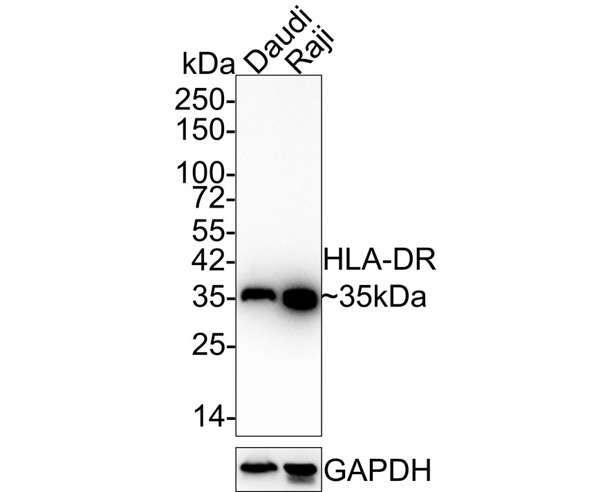
Western blot analysis of HLA-DR on different lysates with Rabbit anti-HLA-DR antibody at 1/1,000 dilution. Lysates/proteins at 20 µg/Lane. Exposure time: 1 minute 40 seconds; 4-20% SDS-PAGE gel. Proteins were transferred to a PVDF membrane and blocked with 5% NFDM/TBST for 1 hour at room temperature. The primary antibody at 1/1,000 dilution was used in 5% NFDM/TBST at 4℃ overnight. Goat Anti-Rabbit IgG - HRP Secondary Antibody at 1:100,000 dilution was used for 1 hour at room temperature.IHC
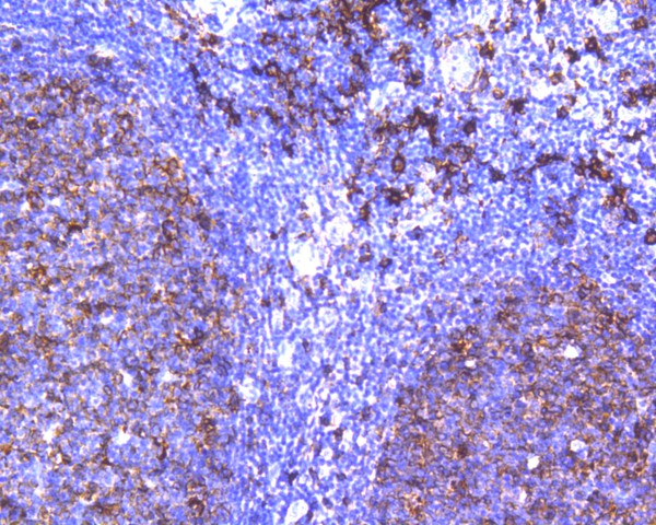
Immunohistochemical analysis of paraffin-embedded human tonsil tissue using anti-HLA-DR antibody. The section was pre-treated using heat mediated antigen retrieval with Tris-EDTA buffer (pH 9.0) for 20 minutes.The tissues were blocked in 5% BSA for 30 minutes at room temperature, washed with ddH2O and PBS, and then probed with the primary antibody (1/200) for 30 minutes at room temperature. The detection was performed using an HRP conjugated compact polymer system. DAB was used as the chromogen. Tissues were counterstained with hematoxylin and mounted with DPX.ICC/IF
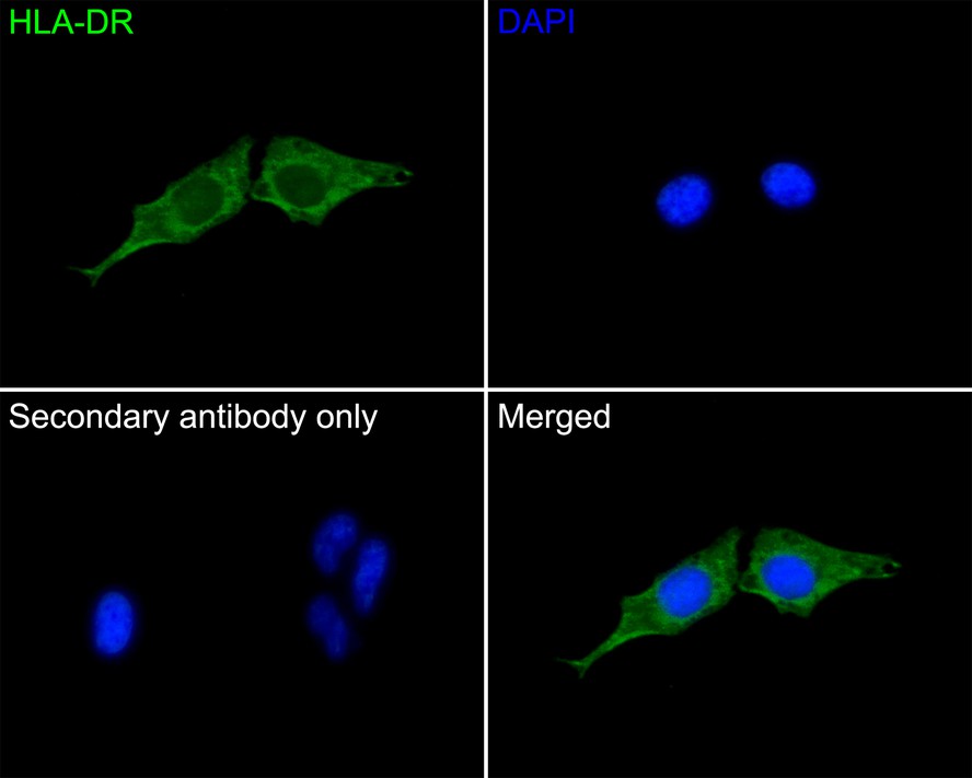
Immunocytochemistry analysis of Hela cells labeling HLA-DR with Rabbit anti-HLA-DR antibody at 1/50 dilution. Cells were fixed in 4% paraformaldehyde for 10 minutes at 37 ℃, permeabilized with 0.05% Triton X-100 in PBS for 20 minutes, and then blocked with 2% negative goat serum for 30 minutes at room temperature. Cells were then incubated with Rabbit anti-HLA-DR antibody at 1/50 dilution in 2% negative goat serum overnight at 4 ℃. Goat Anti-Rabbit IgG H&L (488) was used as the secondary antibody at 1/1,000 dilution. PBS instead of the primary antibody was used as the secondary antibody only control. Nuclear DNA was labelled in blue with DAPI.| Product Name | HLA-DR Recombinant Rabbit Monoclonal Antibody |
|---|---|
| Antibody Type | Primary Antibodies |
| Immunogen | Synthetic peptide within Human HLA-DRA aa 159-195 (Extracellular). |
| Clonality | Monoclonal |
|---|---|
| Isotype | IgG |
| Host Species | Rabbit |
| Tested Applications | ICC/IFIHCWB |
| WB:1:1000 IHC:1:50-1:200 ICC/IF:1:50-1:200 |
|
| Species Reactivity | HumanMouse |
| Concentration | 1mg/ml |
| Purification | Protein A |
| Gene Symbol | HLA-DRA |
|---|---|
| Gene Synonyms | HLA-DRA1 |
| Gene Full Name | major histocompatibility complex, class II, DR alpha |
| Gene Summary | HLA-DRA is one of the HLA class II alpha chain paralogues. This class II molecule is a heterodimer consisting of an alpha and a beta chain, both anchored in the membrane. This molecule is expressed on the surface of various antigen presenting cells such as B lymphocytes, dendritic cells, and monocytes/macrophages, and plays a central role in the immune system and response by presenting peptides derived from extracellular proteins, in particular, pathogen-derived peptides to T cells. The alpha chain is approximately 33-35 kDa and its gene contains 5 exons. Exon 1 encodes the leader peptide, exons 2 and 3 encode the two extracellular domains, and exon 4 encodes the transmembrane domain and the cytoplasmic tail. DRA does not have polymorphisms in the peptide binding part and acts as the sole alpha chain for DRB1, DRB3, DRB4 and DRB5. [provided by RefSeq, Aug 2020] |
| Alternative Names | DR alpha chain antibody DR alpha chain precursor antibody DRA_HUMAN antibody DRB1 antibody DRB4 antibody Histocompatibility antigen HLA DR alpha antibody HLA class II histocompatibility antigen antibody HLA class II histocompatibility antigen DR alpha chain antibody HLA DR1B antibody HLA DR3B antibody HLA DRA antibody HLA DRA1 antibody HLA DRB1 antibody HLA DRB3 antibody HLA DRB4 antibody HLA DRB5 antibody HLA-DRA antibody HLADR4B antibody HLADRA1 antibody HLADRB antibody Major histocompatibility complex class II DR alpha antibody Major histocompatibility complex class II DR beta 1 antibody Major histocompatibility complex class II DR beta 3 antibody Major histocompatibility complex class II DR beta 4 antibody Major histocompatibility complex class II DR beta 5 antibody MGC117330 antibody MHC cell surface glycoprotein antibody MHC class II antigen DRA antibody MHC II antibody MLRW antibody |
| Molecular Weight(MW) | 29 kDa(Observed band size:35 kDa) |
| Cellular Localization | Cell membrane, Endoplasmic reticulum membrane, Golgi apparatus, Endosome membrane, Lysosome membrane. |
WB
Western blot analysis of HLA-DR on different lysates with Rabbit anti-HLA-DR antibody at 1/1,000 dilution. Lysates/proteins at 20 µg/Lane. Exposure time: 1 minute 40 seconds; 4-20% SDS-PAGE gel. Proteins were transferred to a PVDF membrane and blocked with 5% NFDM/TBST for 1 hour at room temperature. The primary antibody at 1/1,000 dilution was used in 5% NFDM/TBST at 4℃ overnight. Goat Anti-Rabbit IgG - HRP Secondary Antibody at 1:100,000 dilution was used for 1 hour at room temperature.
IHC
Immunohistochemical analysis of paraffin-embedded human tonsil tissue using anti-HLA-DR antibody. The section was pre-treated using heat mediated antigen retrieval with Tris-EDTA buffer (pH 9.0) for 20 minutes.The tissues were blocked in 5% BSA for 30 minutes at room temperature, washed with ddH2O and PBS, and then probed with the primary antibody (1/200) for 30 minutes at room temperature. The detection was performed using an HRP conjugated compact polymer system. DAB was used as the chromogen. Tissues were counterstained with hematoxylin and mounted with DPX.
ICC/IF
Immunocytochemistry analysis of Hela cells labeling HLA-DR with Rabbit anti-HLA-DR antibody at 1/50 dilution. Cells were fixed in 4% paraformaldehyde for 10 minutes at 37 ℃, permeabilized with 0.05% Triton X-100 in PBS for 20 minutes, and then blocked with 2% negative goat serum for 30 minutes at room temperature. Cells were then incubated with Rabbit anti-HLA-DR antibody at 1/50 dilution in 2% negative goat serum overnight at 4 ℃. Goat Anti-Rabbit IgG H&L (488) was used as the secondary antibody at 1/1,000 dilution. PBS instead of the primary antibody was used as the secondary antibody only control. Nuclear DNA was labelled in blue with DAPI.| Application Notes | WB:1:1000 IHC:1:50-1:200 ICC/IF:1:50-1:200 |
|---|
| Form | Liquid |
|---|---|
| Storage Instructions | Shipped at 4℃. Store at +4℃ short term (1-2 weeks). Store at -20℃ long term. |
| Storage Buffer | 1*PBS (pH7.4), 0.1% BSA, 40% Glycerol, 0.2% Proclean 950. |


